Download
1 / 11
121 likes | 363 Vues
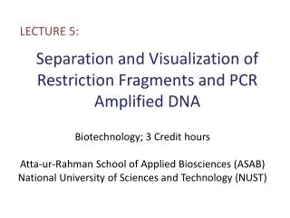
LECTURE 5:. Separation and Visualization of Restriction Fragments and PCR Amplified DNA. Biotechnology; 3 Credit hours Atta-ur-Rahman School of Applied Biosciences (ASAB) National University of Sciences and Technology (NUST). Gel Electrophoresis.

E N D
LECTURE 5: Separation and Visualization of Restriction Fragments and PCR Amplified DNA Biotechnology; 3 Credit hours Atta-ur-Rahman School of Applied Biosciences (ASAB) National University of Sciences and Technology (NUST)
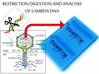
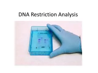
Gel Electrophoresis • Restriction enzyme digestions and other manipulations of DNA enable the results to be directly visualized. • Gel electrophoresis is a technique for separating DNA fragments by size and visualizing them after staining. • It separates molecules on the basis of their rate of movement through a gel under the influence of an electrical field • Gelatinous agarose is used as the medium through which DNA passes • Ethidium bromide is used for staining
- + Power • DNA is negatively charged. • When placed in an electrical field, DNA will migrate toward the positive pole (anode). • An agarose gel is used to slow the movement of DNA and separate by size. - +
DNA - + Power How fast will the DNA migrate? • Strength of the electrical field, buffer, density of agarose gel • Size of the DNA • Small DNA move faster than large DNA • Gel electrophoresis separates DNA according to size small large Within an agarose gel, linear DNA migrate inversely proportional to the log10 of their molecular weight.
Agarose is a linear polymer extracted from seaweed D-galactose 3,6-anhydro L-galactose • Agarose is a polysaccharide obtained from agar. • Agar is a gelatinous substance derivedfrom a polysaccharide in red algae, where it accumulates in the cell walls of agarophyte and serve as the primary structural support for the algae's cell walls • Agarose forms an inert matrix utilized in separation techniques.
Polymerized agarose is porous, allowing for the movement of DNA Scanning Electron Micrograph of Agarose Gel (1×1 µm) Agarose Gel
12,000 bp 5,000 DNA migration 2,000 1,650 1,000 850 650 500 400 300 200 100 DNA Ladder Standard - bromophenol blue + Inclusion of a DNA ladder (DNAs of know sizes) on the gel makes it easy to determine the sizes of unknown DNAs. Note: bromophenol blue migrates at approximately the same rate as a 300 bp DNA molecule
Staining of Agarose Gel • DNA by itself is not visible in gel • Ethidium bromide is usually added to make the DNA band visible • Ethidium bromide molecule interclate between the bases causing the DNA to fluorescence orange when the gel is illuminated with UV light • Methylene blue is also used • Most labs use ethidium bromide as it is very sensitive
Ethidium Bromide Ethidium binds by inserting itself bewteen the stacked bases in double-stranded DNA. The ring structure of ethidium is hydrophobic and resembles the rings of the bases in DNA. Ethidium is capable of forming close van der Walls contacts with the base pairs and that's why it binds to the hydrophobic interior of the DNA molecule.