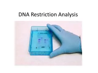
RESTRICTION DIGESTION AND ANALYSIS OF LAMBDA DNA
340 likes | 1.11k Vues
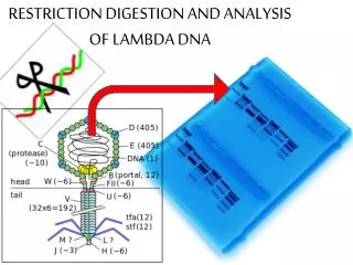
RESTRICTION DIGESTION AND ANALYSIS OF LAMBDA DNA. SAFETY FIRST. WEAR GLOVES WASH HANDS WITH SOAP WHEN DONE HAPPY BIRTHDAY. JUMP TO SLIDE 22. What was the point of this lab?. What is lambda( λ ) ? Why lambda?. Lambda is a bacteriophage , virus that infects bacteria

RESTRICTION DIGESTION AND ANALYSIS OF LAMBDA DNA
E N D
Presentation Transcript
SAFETY FIRST • WEAR GLOVES • WASH HANDS WITH SOAP WHEN DONE • HAPPY BIRTHDAY JUMP TO SLIDE 22
What is lambda(λ)? Why lambda? • Lambda is a bacteriophage, virus that infects bacteria • Inserts its nucleic acid into the host bacterial cell • Replicates rapidly inside host cells until the cells burst and release more phages • Harmless to man and other eukaryotic organisms SOOOO, excellent source of DNA for experimental study.
Lambda genome is about 48,000 bp If linear lambda DNA is cut with HindIII, how many fragments will there be? Longest piece? Shortest piece?
What is a restriction enzyme? • Enzymes that cut DNA at specific places known as restriction sites • Also called endonucleases • Bacteria use them as a natural defense against bacteriophages • Biotechnology – cutting genes from one organism and pasting them into another – would not be possible without these enzymes
Enzyme Site Recognition Restriction site Palindrone • Each enzyme digests (cuts) DNA at a specific sequence = restriction site • Enzymes recognize 4- or 6- base pair, palindromic sequences (eg GAATTC) Fragment 2 Fragment 1
5 vs 3 Prime Overhang Enzyme cuts • • Generates 5 prime overhang • DNA from any organism cut with the same enzyme will produce complementary sticky ends • When mixed together, complementary bases will hydrogen bond • Ligase is needed to reform the phosphodiester bonds.
5’GAATTC3’ 3’CTTAAG5’ 5’AAGCTT3’ 3’TTCGAA5’ 5’CTGCAG3’ 3’CACGTC5’ Common Restriction Enzymes • EcoRI • Escherichiacoli • 5’ overhang • HindIII • Haemophilusinfluensae • 5’ overhang • PstI • Providenciastuartii • 3’ overhang
How is a restriction digest done? • Restriction Buffer provides optimal conditions for enzyme • Why incubate at 37°C? • Body temperature is optimal for these and most other enzymes • What happens if the temperature is too hot or cool? Too hot = enzyme may be denatured Too cool = enzyme activity lowered, requiring longer digestion time
How can we separate all those fragments of DNA? • Agarose gel electrophoresis • Agarose is purified agar • Derived from seaweed • agar provides a medium on which bacteria (and other microorganisms can grow) • agarose provides a “sieve” for separating DNA fragments by size • Large fragments travel slower than small fragments
Gel electrophoresis • DNA is negatively charged • when it’s in an electrical field it moves toward the positive side DNA – + “swimming through Jello”
AgaroseElectrophoresisLoading • Electrical current carries negatively-charged DNA through gel towards positive (red) electrode Buffer Dyes Agarose gel Power Supply
How can we see the DNA fragments – DNA is not colored? • Stain them! • The stain we use is relatively nontoxic, easy to handle and dispose of • BUT it’s not very sensitive so in “real life” other types of stains are used • Loading dyes and tracking dyes do not stain DNA; they just help you see where your sample is and how far DNA fragments have probably traveled
How can we determine the sizes of the DNA fragments? • Run a standard in one of the wells; also called marker or ladder • Standard has been cut with restriction enzymes and the size in base pairs (bp) has been determined • Compare migration of fragments whose size is known to the migration of fragments whose size is unknown
How can we determine the sizes of the DNA fragments? • Create a standard curve using the migration of the marker DNA fragments • Determine the size of the unknown fragments from this graph • Semi-log graph paper will be needed
Analysis of Stained Gel Determine restriction fragment sizes • Create standard curve using DNA marker • Measure distance traveled by restriction fragments • Determine size of DNA fragments • Identify the related samples
Molecular Weight Determination Fingerprinting Standard Curve: Semi-log Size (bp) Distance (mm) 23,000* 11.0 9,400 13.0 6,500 15.0 4,400 18.0 2,300 23.0 2,000 24.0 *This fragment falls outside the linear portion of the curve. You may choose to exclude it from your best fit line
OVERVIEW • Page 21 You can answer (3 questions) • Page 22 You can answer (4 questions) • Page 23 You can answer (2 questions)
LESSON 1 • Page 24 Let’s answer the two questions in the middle of the page • Be sure to fill out the chart • Page 25You can answer – 2 questions at the top and the 4 review questions at the bottom • Page 26 You can answer – 4 questions • Page 27 You can answer – 4 questions
LESSON 2 • Page 30 You can answer – 5 questions • Page 31 You can answer – 2 questions • Page 32 At the top – Let’s answer that now
LESSON 3 • follow procedure 2. a on page 35 • Page 36 Do not attach your tracing here; hand in separately; if you have no data another group will share with you – be sure to document where the data came from • Page 37; if you followed directions the wells are in this order: Lane 1 Marker (we know the size of these fragments; they were cut with HindIII ) Lane 2 Uncut lambda Lane 3 lambda cut with PstI Lane 4 lambda cut with EcoRI Lane 5 lambda cut with HindIII
LESSON 3 • Measure from the front of the well to the front of the band • RECORD DATA ON PAGE 38 • BE CAREFUL GEL IS FRAGILE!
LESSON 3 • Page 39 You do – 6 questions • Page 40 You do – 2 questions • Page 41 You do – 1 question • Clarification step 3 use marker • Page 42 – Complete the graph – you may ignore the 23,000 bp piece when drawing the best fit line • Page 43 in the “estimated” columns also include the data from the first gel analysis in parenthesis; the number from the standard curve graph should not be in parenthesis • Page 44 You do – 4 questions
Lane 1: marker, lambda cut with HindIII • Lane 2: uncut lambda • Lane 3: lambda cut with PstI • Lane 4: lambda cut with EcoRI • Lane 5: lambda cut with HindIII
Lane 1: marker, lambda cut with HindIII; 7 sites, 8 pieces • Lane 2: uncut lambda; 48,502 bp • Lane 3: lambda cut with PstI; 28 sites, 29 fragments • Lane 4: lambda cut with EcoRI; 5 sites, 6 fragments • Lane 5: lambda cut with HindIII
WHY DIDN’T WE SEE ALL THE FRAGMENTS AS BANDS IN THE GEL? • SOME BANDS ARE SO CLOSE IN SIZE THEY DID NOT SEPARATE USING THIS PROTOCOL • SOME FRAGMENTS ARE SO SMALL THEY CAN NOT BE DETECTED • How could we get better results? • Change gel concentration • Longer run time • More sensitive DNA stain
EXPERIMENTAL ERROR • *Incorrect measurement • *Not using optimal temperature for enzyme for the right amount of time • *Some of sample did not enter well • *Stock solutions not kept on ice