Download
1 / 41
410 likes | 438 Vues
Understand restriction enzymes, analyze DNA fragments, and learn Southern blotting techniques in this comprehensive experiment guide. Discover the role of restriction enzymes in microorganisms and their applications in recombinant DNA technology. Explore different types of restriction enzymes and the unit determination assay. Enhance your knowledge of Southern blotting, a pivotal molecular biology technique developed by E.M. Southern in 1975.

E N D
Experiment Goals • Digestion of DNA by restriction enzyme • Analyze digested DNA by electrophoresis • Transfer digested DNA to nitrocellulose filters (Southern blotting) • Procedure of setting up a Southern blotting
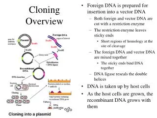
Restriction Enzymes • Definition: • A restriction enzyme (or restriction endonuclease) is an enzyme that cuts double-stranded DNA at specific sites that it recognizes. • The enzyme makes two incisions, one through each of the sugar-phosphate backbones (i.e., each strand) of the double helix without damaging the nitrogenous bases.
Restriction enzyme The enzyme EcoRI cutting DNA at its recognition sequence • Different restriction enzymes have different recognition sequences. • This makes it possible to create a wide variety of different gene fragments.
Function of Restriction Enzymes in microorganisms • Provide microorganisms with resistance to invading organisms or foreign DNA. • Endonucleases in bacterial cells resist infections by viruses, by destroying foreign DNA molecules. • consist of a related pair of enzymes • Endonuclease – cuts foreign DNA • Methylase – protects host DNA
Methylase Enzymes • Restriction enzymes usually occur in combination with one or two modification enzymes (DNA-methyltransferases) • Protect the cell’s own DNA from cleavage by the restriction enzyme. • Modification enzymes recognize the same DNA sequence as the restriction enzyme that they accompany, • Instead of cleaving the sequence, they methylate one of the bases in each of the DNA strands. • The methyl groups protrude into the major groove of DNA at the binding site and prevent the restriction enzyme from acting upon it.
Naming • Restriction enzymes are named based on the bacteria in which they are isolated in the following manner: example “EcoRI” • E Escherichia (genus) • co coli (species) • R RY13(strain) • I First identified Order
Restriction Enzyme • There are hundreds of different REs from different microorganisms • Each RE cuts DNA at a specific “recognition sequence” of nucleotides. Examples: • EcoRI-- GAATTC; • AluI -- AGCT • Each recognizes its specific “recognition sequence” and cuts both strands of DNA wherever that sequence is found, but nowhere else.
Restriction Enzyme Uses • Recombinant DNA technology • Cloning • Replicates a sequence inserted into a host cell • DNA restriction mapping • A rough map of a DNA fragment • DNA fingerprints
Types of Restriction Enzymes • Restriction enzymes are traditionally classified into three types on the basis of • subunit composition, • cleavage position, • sequence-specificity • and cofactor-requirements
Types of Restriction Enzymes • Type I - Recognize specific sequences and cut DNA at a nonspecific site > than 1,000 bp away • Type II - Recognize palindromic sequences and cut within the palindrome • Type III - Recognize specific 5-7 bp sequences and cut 24-27 bp down stream of the site. • Type II restriction enzymes are the most useful class as they recognize specific palindomic sequences in DNA and cut the sugar phosphate backbone within the palindrome
Restriction Enzymes and DNA fragments • A restriction enzyme functions by "scanning" the length of a DNA molecule. • Once it encounters its particular specific recognition sequence, • it will bind to the DNA molecule • and makes one cut in each of the two sugar-phosphate backbones of the double helix.
Endonucleases and DNA fragments • Blunt ends • Sticky ends
Unit Determination Assay • One unit of restriction endonuclease is defined as the amount of enzyme required to digest one microgram of the appropriate substrate DNA completely in 60 minutes under the conditions specified for that enzyme.
Set up of a restriction enzyme reaction • A RE reaction contains the DNA to be analyzed, • A restriction enzyme, • A restriction enzyme buffer mix. • contains a buffering agent to maintain constant pH, • and Mg++ (from MgCl2) as a necessary cofactor for enzyme activity.
HinfI Restriction Enzyme • Recognition Site:
Electrophoresis of Genomic DNA Odd numbered lanes contain undigested genomic DNA Even numbered lanes contain digested genomic DNA
Southern Blotting • The technique was developed by E.M. Southern in 1975.
What Is Southern Blotting? • A technique used in molecular biology to check for the presence of a particular DNA sequence in a DNA sample.
Southern Blot • The Southern Blot takes advantage of the fact that DNA fragments will stick to a nylon or nitrocellulose membrane. • The membrane is laid on top of the agarose gel and absorbent material (e.g. paper towels or a sponge) is placed on top. • With time, the DNA fragments will travel from the gel to the membrane by capillary action as surrounding liquid is drawn up to the absorbent material on top. • The membrane is now a mirror image of the agarose gel.
Uses of Southern Blotting • Identify mutations, deletions, and gene rearrangements • Used in prognosis of cancer and in prenatal diagnosis of genetic diseases • diagnosis of Leukemias • detect variations in gene structure • identify homologous genes among different species
Performing Southern Blotting • DNA Digestion with an appropriate restriction enzyme. • Gel Electrophoresisrun the digest on an agarose gel. • Denature the DNA (usually while it is still on the gel). • Transfer the denatured DNA to the membrane (blotting) • Preparing the probe • HybridizationProbe the membrane with labeled ssDNA. • DetectionVisualizeyour radioactively labeled target sequence.
1- DNA Digestion • Cut the DNA into different sized pieces. • HinfI restriction enzyme is used
2- Gel Electrophoresis • Sorts the DNA pieces by size • Agarose or polyacrimide
Electrophoresis of Genomic DNA Odd numbered lanes contain undigested genomic DNA Even numbered lanes contain digested genomic DNA
3- Denature the DNA • DNA is then denatured with an alkaline solution such as NAOH. • This causes the double stranded to become single-stranded.
4- Blotting • Transfer the DNA from the gel to a solid support. • The blot is usually done on a sheet of nitrocellulose paper or nylon. • Transferred by either electrophoresis or capillary blotting.
4- Blotting 1) Electrophoresis- takes advantage of the molecules negative charge.
Agar gel with DNA Weight Wick (filter paper) Filter paper Buffer Membrane 4- Blotting 2) Capillary blotting-fragments are eluted from the gel and deposited onto the membrane by buffer that is drawn through the gel by capillary action. Paper towel stack
4- Blot Fixation • The blot is made permanent by: • Drying at ~80°C • Exposing to UV irradiation
5- Preparing the probe • It is a fragment of DNA of variable length (usually 100-1000 bases long), which is used to detect in DNA the presence of nucleotide sequences that are complementary to the sequence in the probe • Must be labeled to be visualized • Usually prepared by making a radioactive copy of a DNA fragment. Probing is often done with 32P labeled ATP, biotin/streptavidin or a bioluminescent probe.
6- Hybridization • Hybridization-process of forming a double-stranded DNA molecule between a single-stranded DNA probe and a single-stranded target patient DNA.
6- Hybridization • Steps for hybridization 1. The labeled probe is added to the matrix incubated for several hours to allow the probe molecules to find their targets 2. Any unbound probes are then removed. 3. The place where the probe is connected corresponds to the location of the immobilized target molecule.
probes 3’ – *ATCTCGGGAATC – 5’ add probe hybridization 5’ – …AAGCCTAGAGCCCTTAGCCAAAAG… – 3’ *ATCTCGGGAATC
7- Detection Visualize your labeled target sequence. • If radiolabeled 32P probe is used, then you would visualize by autoradiography. • Biotin/streptavidin detection is done by colorimetric methods, • and bioluminescent visualization uses luminescence.
Steps in Southern Blotting DNA extraction Disease gene Fragments of DNA appear as a smear DNA digestion Gel electrophoresis Paper towels Denaturation of patient’s DNA in gel Chromatography paper support Gel in NaOH Nylon filter Southern blot Gel 10x SSC Blot dismantled Autoradiography X ray film Hybridisation: Stringency washes Radioactive probe added to filter cassette filter filter