Download
1 / 118
1.24k likes | 2.76k Vues
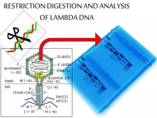
Restriction Enzyme digestion of DNA - Exercise 8. Objectives -Understand how Restriction Enzymes digest DNA. -Know how to construct a pAMP (plasmid) or gel. -Given the size of fragments, gel, know how to construct a restriction map. -Given a restriction map know how to construct a gel.

E N D
Restriction Enzyme digestion of DNA - Exercise 8 Objectives -Understand how Restriction Enzymes digest DNA. -Know how to construct a pAMP(plasmid) or gel. -Given the size of fragments, gel, know how to construct a restriction map. -Given a restriction map know how to construct a gel.
NOTE: DNA IS Negatively charge because of the phosphate groups.
DNA molecules are macromolecules that hold the genetic information of living organisms. They are extremely long, double-stranded polymers of nucleotides. • The covalent bond joining adjacent nucleotides in DNA is called a phoshodiester bond. • The phoshodiester bonds between nucleotides in DNA molecules are very stable unless they are physically stretched or exposed to enzymes name nucleases.
Enzymes are capable of breaking (hydrolyzing) phoshodiester bonds in DNA molecules. Nucleases can be classified into two major groups: exonucleases and endonuclases. • Exonucleases: If the enzyme digest nucleotides from the ends of the DNA molecules. • Endonuclases: If the enzyme digest nucleotides in the interior of a DNA molecule. • Restriction endonuclease: An enzythem that digest DNA by recognizing specific short sequences of bases that are called palindromes.
A special class of endonucleases from a bacteria has been isolated for this experiment. These special enzymes, termed restriction endonucleases (RE),digest DNA by breaking bonds only within a specific short sequence of bases. These base sequences usually ran in size from 4-8 base pairs but can be as long as 23 base pairs. • Restriction endonucleases confer an adaptive advantage on bacteria by digesting foreign DNA usually from an invading bacteriphage (bacterial virus). The resulting DNA fragments can then be further degraded and destroyed by exonucleases. These enzymes are used to cut DNA in a precise and predictable manner. They are extensively useful in gene cloning, DNA amplification, and many recombinant DNA technologies. • Restriction endonuclease (RE). This RE are also attained from bacteria. In a bacteria where we get these enzymes form there protected because if a virus invades a bacteria cell these endonuclease will chop up the virus DNA, its like a defense system, so we can isolate these endonuclease for experiments, but bacteria produce these endonuclease to protect themselves from foreign DNA entering their cells.
2 Restriction Endonucleases (RE) • EcoR1 & HindIII. Both of these recognize different nucleotide sequences. • Each strand of DNA is cut at the phoshodiester bond between the G and A bases (indicated by the arrow signs). Notice that the sequence GAATTC is the same on both strands when each strand is read 5’ -> 3’. Such symmetrical sequences are called palindromes (In a English language a palindrome reads the same thing in both directions). This enzyme cuts the double strands asymmetrically, leaving protruding ends. These protruding bases are referred to as sticky ends aka compatible cohesive ends.
EcoR1: EcoR1 recognizes palindrome on DNA, and cuts the bond between G & A, and G & A. When you do that it opens your DNA. For example, if you have plasmid and that palindrome is present once on the plasmid, you’ll get one cut. • If somewhere else that palindrome is present and you incubate it with EcoR1, you’ll get another cut. So every time EcoR1 recognizes this palindrome on your plasmid, it will cut through the DNA. So when it opens up the DNA, may get a couple of unpaired bases, and those unpaired bases are called sticky ends, and if you throw some nucleotides from different species, you can make recombinant DNA.
Like EcoRI, HindIII also recognizes a palindromic sequence, AAGCTT, and produces sticky ends. Sticky ends can hydrogen bond together other because of complementary base pairing.
Recombinant DNA molecules are compose of DNA fragments from two or more sources. Not all RE’s produce sticky ends. Some enzymes cut DNA to produce blunt ends, as shown here.
Once the DNA has been digested, the fragments must be separated and identified. Fragments are separated by agarose gel electrophoresis. Agar is a large polysaccharide. • Gel electrophoresis: You put an agorose gel (agrose is a polysaccharide) and it has spaces, your DNA can move through these spaces, you put a current against this, the negative end is up, the positive is at the bottom, and because your DNA has a negative charge, the DNA moves down towards the positive end. • Gel is immersed in an ionic buffer. The buffer has a pH above 8.0 DNA at this pH is negatively charge because the phosphates in the DNA backbone have lost hydrogen ions. The dye molecules serve as the indicator of the movements of invisible DNA molecule through the gel as an electric current is run through the gel. The negatively charged DNA will migrate from the anode to the cathode (negative to positive) along with the current.
Separation of the DNA fragments occurs as they migrate through the network of agarose molecules. Smaller fragments slip through the network fast than large molecules. The rate of migration is a function of fragment size, as well as the density of agarose. The tightness (concentration of agarose). • High concentration favor smaller fragments. • Low concentration favor large fragments. • Each of migration function of fragment size and density of agarose. Depending on what conformation a circular DNA gets, it will run differently in the gel. So not only does the size of the DNA molecule affect migration rate, but the configuration of the DNA also affects the migration rate. The DNA that you will electrophoresing can exist in three different conformations.
1. Supercoil circular: 1st fastest. When its circular it becomes twisted and turn and be comes a little bit shorter in size. Migrates fastest down the gel. Contains small volume, more compacted. • 2. Linear: Migrates next fastest down the gel. • 3. Nicked (relaxed) circular: One strand is intact, the other is broken and when it is nicked, it becomes extended. This one is very relaxed and faces the most difficulty making its way through the agarose. Supercoil < Linear < Nicked (relaxed) circular
In addition to conformation affecting migration rate, laboratory production of plasmid DNA can be produce very large molecules that migrate very slowly. Two possible molecules that can be produced are dimers and concatemers. A dimer consists of two plasmids covalently linked in a series end to end. A Concatemer, for example, might consist of two plasmids with one hooked through the other but not covalently linked to each other. If a purified uncut plasmid is applied to a gel, bands of super coiled plasmid, nicked circular plasmid, dimers, and concatemer can be observed. • Dimer: Means that its link together by 2 links • Concatemer: Mean a whole bunch of plasmids linked together but not covalently linked to each other.
pAMP - the plasmid DNA. What we did in the experiment on DNA restriction analysis is we took pAMP (circle) and incubated the pAMP this plasmid with different restriction endonucleases. • From your electrophoresis gel, you can estimate the size of pAMP. You can also determine if pAMP is circular or linear. Finally, you can use the gel to draw a restriction map. A restriction map is a physical map of a piece of DNA showing recognition sites of specific restriction enzymes separated by lengths marked in numbers of bases. Separated DNA base on size • The pattern of DNA bands is characteristic for a specific DNA sample and the restriction enzymes used to cleave it. A banding pattern can be referred to as a DNA fingerprint. because it is unique to that particular DNA (and the combination of restriction fragments). • We ran a gel to see if we could determine how many DNA fragments you got. By electrophoresing a series of fragments of known size (DNA ladder) along with the DNA samples of interest, the sizes of unknown fragments can be estimated.
A restriction site is a place where an enzymes cuts DNA, so there are restriction sites for EcoR1, and for HindIII. • When constructing the pAMP no restriction site where you start and where you finish. • Lane 4: A control to see what uncut plasmid looks like. How uncut DNA traveled whether they made 1 or 2 pieces. It’s your plasmid DNA DNA was on tube 4 which acts like a measurements and acts like a ladder. No enzyme (Lane 4). • Lane 5: DNA ladder: DNA digest, containing known base pair lengths compare with fragments in lanes 1-3. You will run DNAs of known size (DNA ladder) to help you estimate the size of your DNA fragments. Lane 5 contains DNAs of known sizes (DNA ladder).
Prokaryotic (Circular) DNA • DNA from bacteria (both chromosomal DNA and extra chromosomal plasmid DNA) and viruses is often a closed circle. If you have a circular DNA, we know that’s Prokaryotic DNA. In Prokaryotic DNA, the number of fragments will equal the number of restriction sites. Eukaryotic (linear) DNA • If you have one restriction site for an enzyme, you would have 2 fragments, and if you have 2 restriction sites for an enzyme, you would have 3 fragments. In Eukaryotic DNA, the number of fragments is always going to have one more or one less than restriction sites. • In Eukaryotic DNA, there’s no reason to see multiply bands in control lane because Eukaryotic DNA is linear, it doesn’t exist as supercoil, relax, or multimere so this is a hint in lane 4. So when you have Eukaryotic DNA, you will not see multiply bands in the control lane. • Also, just because they show you multiply bands, not every time your going to have prokaryotic (circular) DNA you get multiply lanes, its only if the DNA has been damaged into a supercoil.
Size: small pieces migrate faster, farther than bigger pieces. • Conformation (shape): Comparing 3 pieces of DNA that are the same size. Supercoil < Linear < Nicked (relaxed) circular • Charge: Charge (+,-)DNA is negative because of Phosphate groups (anode) to positive (cathode).
Digestion of pAMP with EcoRI & HindIII • We incubated our plasmid under several conditions. Those conditions were that we incubate pAMP. • Lane 1: EcoR1 - one band • Lane 2: HindIII - one band • Lane 3: EcoR1 & HindIII - two bands • Lane 4: Water - Our control. We got one main band. • Lane 5: DNA ladder, a tool to measure the size of DNA fragments.
When constructing the pAMP. There’s no restriction site where you start and where you finish the map. You could call this point the reference point. Also, all your base pairs (fragments) have to equal the total number base pairs of your plasmid. For example, 6,000 Bp’s in this example.
Key for pAMP KEY Enzyme A: Light green Enzyme B: Pink Enzyme C: Orange