Download
1 / 40
420 likes | 505 Vues
Learn about the pathology, pathogenesis, and complications of atherosclerosis and ischemic heart disease, including myocardial infarction. Understand lifestyle modifications to reduce risks. Study arterial conditions like arteriosclerosis and vasculitis. Explore risk factors, morphological changes, and clinical features of MI.

E N D
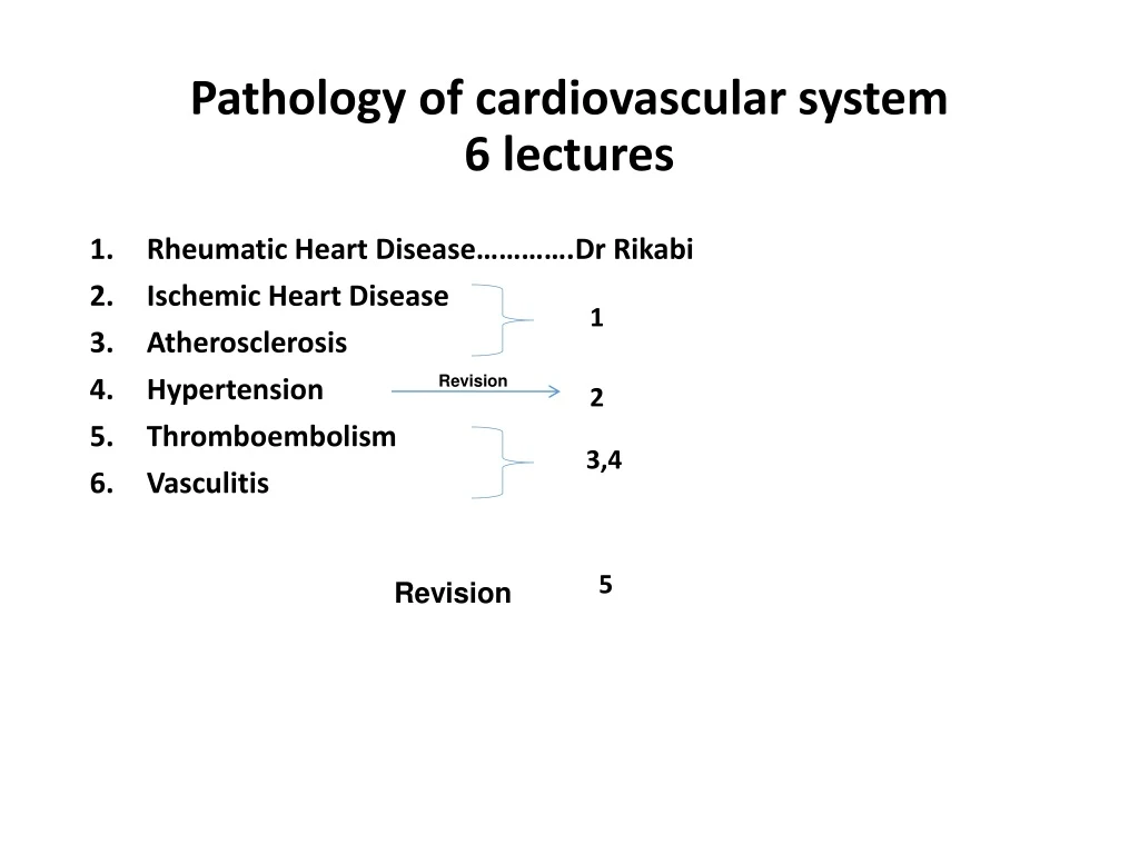
Pathology of cardiovascular system6 lectures • Rheumatic Heart Disease………….Dr Rikabi • Ischemic Heart Disease • Atherosclerosis • Hypertension • Thromboembolism • Vasculitis 1 Revision 2 3,4 5 Revision
Atherosclerosis and ischemic heart diseaseAhmed Alhumidi. Pathology. KSU. Riyadh. 2015
Atherosclerosis and ischemic heart disease Objectives: • Understand the pathology, pathogenesis and clinical consequences of atherosclerosis. • Be able to discuss pathology and complications of ischaemic heart diseases with special emphasis on myocardial infarction. • Know how lifestyle modifications can reduce the risk of ischaemic heart disease.
Arteriosclerosis"hardening of the arteries") is a generic term for thickening and loss of elasticity of arterial walls. Atherosclerosis Mönckeberg medial calcific sclerosis Arteriolosclerosis Old age Hypertention
Atherosclerosis (AS) • Atherosclerosis is characterized by intimal lesions called atheroma, which protrude into and obstruct vascular lumens and weaken the underlying media. Atheroma= Intimal thickening + lipid accumulation
Atheroma Atherosclerotic plaques have three principal components: Cells: SMCs, foam macrophages, lymphocytes Extracellular matrix: collagen, elastic fibers Lipid: Typical atheromas contain relatively abundant lipid both intracellular and extracellular lipid .
COMPLICATIONS Rupture, ulceration Hemorrhage thrombosis, Aneurysmal dilation. fibrous plaques complicated lesions Fatty streaks= macrophage-derived foam cells
Natural history of atherosclerosis Slide 12.5
Risk Factors for Atherosclerosis anti-C. pneumoniae antibodies detection of the organism within atherosclerotic lesions,
Importance of types of lipoproteins in hypelipidemia • Low-density lipoproteins (LDLs) • Very-low-density lipoproteins (VLDLs) • Chylomicrons High-density lipoproteins (HDLs) “good” cholesterol, because high levels seem to protect against heart attack.
Risk Factors PATHOGENESIS
SUMMARY treatment : Angioplasty http://www.pathophys.org/
Ischemic Heart Disease Ahmed Alhumaidi Pathology Department KSU, Riyadh 2015 Reference: Robbins & Cotran Pathology and Rubin’s Pathology
Ischemic Heart Disease atherosclerosis insufficient blood supply Myocardium • Angina pectoris (chest pain). • Acute myocardial infarction. • Sudden cardiac death. • Chronic ischemic heart disease
Pathogenesis • A lesion obstructing 70% to 75% or more of a vessel lumen-so-called critical stenosis-generally causes symptomatic ischemia (angina) only in the setting of increased demand • A fixed 90% stenosis can lead to inadequate coronary blood flow even at rest Rupture, fissuring, or ulceration
Angina pectoris • Angina pectoris is a type of IHD characterized by paroxysmal and usually recurrent attacks of substernal chest discomfort (variously described as constricting, crushing, squeezing, choking, or knifelike). May radiate down the left arm or to the left jaw (referred pain) . Intermittent chest pain caused by transient, reversible ischemia • Typical (stable) angina • pain on exertion • fixed narrowing of coronary artery • Unstable (pre-infarction) angina • increasing pain with less exertion • plaque disruption and thrombosis • Prinzmetal (variant) angina • pain at rest • coronary artery spasm of unknown etiology ذبحة صدرية مخالفة للمعتاد،
Myocardial Infarction • Definition: MI, also known as "heart attack," is the death of cardiac muscle resulting from ischemia. • Risk factors are the same as those of atherosclerosis. Disruption of an atherosclerotic plaque results in the formation of thrombus
A. Plaque rupture without superimposed thrombus in a patient who died suddenly. B. Acute coronary thrombosis superimposed on an atherosclerotic plaque with focal disruption of the fibrous cap, triggering fatal myocardial infarction. C. Massive plaque rupture with superimposed thrombus, also triggering a fatal myocardial infarction (special stain highlighting fibrin in red). In both
Pathogenesis of MI • Myocardial necrosis begins within 20-30 minutes, mostly starting at the subendocardial region (less perfused, high intramural pressure). • Infarct reaches its full size within 3-6 hrs., during this period, lysis of the thrombus by streptokinase or tissue plasminogen activator, may limit the size of the infarct. • Irreversible cell injury: 20-40 min Types • Transmural • Full thickness (>50% of the wall) • Subendocardial • Inner 1/3 of myocardium
day 1, Day 2, Day 3, Day 4, Day 5, Day 6, day 7 Neutrophils Macrophages • Coagulation necrosis MI: day 1, day 3, day 7
2 weeks • 1 month • Granulation tissue collagenous scar
Myocardial Infarction: Clinical Features • Pain: • Severe crushing sub-sternal chest pain, which may radiate to the neck, jaw, epigastrium, shoulder or left arm. • Pain lasts for hours to days and is not relieved by nitroglycerin. • Absent in 20% of patients (diabetics, hypertensive, elderly). • Pulse is rapid and weak. • Diaphoresis (sweating) • Dyspnea. • Cardiogenic shock in massive MI(>40%of lt. ventricle). • ECG shows typical findings of ischemia. shock is low blood perfusion to tissues
Ischemic Heart Disease Laboratory evaluation • Troponins: best marker, TnT, TnI (more specific). • TnI and TnT are not normally detectable in the circulation • After acute MI both troponins become detectable after 2 to 4 hours, peaks at 48 hours. Their levels remain elevated for 7 to 10 days • CK-MB is the second best marker: • It begins to rise within 2 to 4 hours of MI, peaks at 24 to 48 hours and returns to normal within approximately 72 hours • Lactate dehydrogenase (LD)… LD1. • Rise 24 hrs, peaks 72 hrs, persists 72 hrs. Troponins are regulatory proteins ((troponin C, troponin I, and troponin T) that are integral to muscle contraction Creatine kinase (CK) —is an enzyme expressed by various tissues and cell types 3 isoypesCK-MM, CK-BB and CK-MB. [M: musle B brain] A lactate dehydrogenase (LDH or LD) is an enzyme found in nearly all living cells [LDH-1— heart RBC, brain].
Myocardial Infarction: Outcomes or complications • No complications in 10-20%. • 80-90% experience one or more of the following complications: • Cardiac arrhythmia (75-90%). Many patients have conduction disturbances and myocardial irritability following MI, which undoubtedly are responsible for many of the sudden deaths. Sudden coronary death can occur due to ventricular arrhythmia. • Left ventricular failure with mild to severe pulmonary edema (60%). • Cardiogenic shock (10%).
Complications of MI • Myocardial rupture: • Thromboembolism (15-49%). thecombination of a local myocardial abnormality in contractility (causing stasis) with endocardial damage (causing a thrombogenic surface) can foster mural thrombosis and, potentially, thromboembolism • Pericarditis • Ventricular aneurysm in which the ventricle is dilated and the wall is thinned out. • External rupture of the infarct with associated bleeding into the pericardial space (hemopericardium). • Progressive late heart failure in the form of chronic IHD.
Myocardial Infarction (MI), summary • Necrosis of heart muscle caused by ischemia • Most due to acute coronary artery thrombosis • irreversible injury/cell death in 20-40 minutes • Prompt reperfusion can salvage myocardium Salvage ينقذ
Myocardial Infarction (MI), summary • Clinical features • Severe, crushing chest pain ± radiation • Not relieved by nitroglycerin, rest • Sweating, nausea, dyspnea • Sometimes no symptoms • Laboratory evaluation • Troponinsdetected within 2-4 hours, remain elevated for a week. • CK-MB increases within 2-4 hours, returns to normal within 72 hours. • Complications • contractile dysfunction • arrhythmias • rupture • chronic progressive heart failure • Prognosis • depends on remaining function and perfusion • overall 1 year mortality: 30% • 3-4% mortality per year thereafter
Chronic ischemic heart disease • Progressive heart failure due to ischemic injury, either from: • prior infarction(s) (most common) • chronic low-grade ischemia
Ischemic Heart Disease Sudden cardiac death • Unexpected death from cardiac causes either without symptoms or within 1 to 24 hours of symptom onset • Results from a fatal arrhythmia, most commonly in patients with severe coronary artery disease
Objectives: Atherosclerosis and ischemic heart disease Understand the pathology, pathogenesis and clinical consequences of atherosclerosis. pathologyCells, Extracellular matrix:, Lipid, fibrous cap Pathogenesis endothelial cell injury….dysfunction….smooth muscle immigration ……macrophages and smooth muscle engulf fat …… smooth muscle proliferation and collagen deposition Complications Arterial obstruction………ischemia Embolizationof plaque material…… ischemia Weakening with rupture of the arterial wall …..aneurism and rupture of vessel Be able to discuss pathology and complications of ischaemic heart diseases with special emphasis on myocardial infarction. Pathology MI Complications MI • how lifestyle modifications can reduce the risk of atherosclerosis and ischaemicheart disease. 0-4 h No pathological findings 4-12h coagulation necrosis 12-1day neutrophils 1 day – 7 days less neutrophils and more macrophages 1 week-2 weeks granulation tissue 2 weeks – 8 weeks fibrosis Typical Angina Unstable angina Prinzmetal angina Arrhythmia Left ventricular failure Cardiogenic shock Myocardial rupture Thromboembolism Serum Troponins and CK MB ECG finding
A 68-year-old obese woman (BMI = 32 kg/m2) presents with substernalchest pain. Results of laboratory studies include an elevated WBC count (13,000/μL), CK-MB of 6.6 ng/mL, and troponin-I of 2.5 ng/mL. ECG confirms a myocardial infarction of the left ventricular wall. Which of the following mechanisms is most likely responsible for the myocardial infarction in this patient? (A) Coronary artery thrombosis (B) Coronary artery vasospasm (C) Decreased collateral blood flow (D) Deep venous thrombosis (E) Paradoxical embolism Ahmed alhumaidi Associate professor
The answer is A: Coronary artery thrombosis. Coronary artery thrombosis is the most common cause of acute myocardial infarction and is often secondary to rupture of an atherosclerotic plaque. • Diagnosis: Myocardial infarction Ahmed alhumaidi Associate professor
A study of atheroma formation leading to atherosclerotic complications evaluates potential risk factors for relevance in a population. Three factors are found to play a significant role in the causation of atherosclerosis: smoking, hypertension, and hypercholesterolemia. These factors are analyzed for their relationship to experimental models for atherogenesis. Which of the following events is the most important direct biologic consequence of these factors? □ (A) Endothelial injury and its sequelae □ (B) Conversion of smooth muscle cells to foam cells □ (C) Alterations of hepatic lipoprotein receptors □ (D) Inhibition of LDL oxidation □ (E) Alterations of endogenous factors regulating vasomotor tone Ahmed alhumaidi Associate professor
(A) Atherosclerosis is thought to result from a form of endothelial injury and the subsequent chronic inflammation and repair of the intima. All risk factors, including smoking, hyperlipidemia, and hypertension, cause biochemical or mechanical injury to the endothelium. Formation of foam cells occurs after the initial endothelial injury. Although lipoprotein receptor alterations can occur in some inherited conditions, these account for only a fraction of cases of atherosclerosis, and other lifestyle conditions do not affect their action. Inhibition of LDL oxidation should diminish atheroma formation. Vasomotor tone does not play a major role in atherogenesis. Ahmed alhumaidi Associate professor
A 59-year-old man has experienced chest pain at rest for the past year. On physical examination, his pulse is 80/min and irregular. The figure shows the microscopic appearance representative of the patient's left anterior descending artery. Which of the following laboratory findings is most likely to have a causal relationship to the process illustrated? • □ (A) Low Lp(a) • □ (B) Positive VDRL • □ (C) Low HDL cholesterol • □ (D) Elevated platelet count • □ (E) Low plasma homocysteine Ahmed alhumaidi Associate professor
(C) The figure shows an arterial lumen that is markedly narrowed by atheromatous plaque complicated by calcification. Hypercholesterolemia with elevated LDL and decreased HDL levels is a key risk factor for atherogenesis. Levels of Lp(a) and homocysteine, if elevated, increase the risk of atherosclerosis. Syphilis (positive VDRL test result) produces endarteritis obliterans of the aortic vasa vasorum, which weakens the wall and predisposes to aneurysms. Although platelets participate in forming atheromatous plaques, their number is not of major importance. Thrombocytosis can result in thrombosis or hemorrhage. Ahmed alhumaidi Associate professor
A 60-year-old man has experienced angina on exertion for the past 6 years. A coronary angiogram performed 2 years ago showed 75% stenosis of the left anterior descending coronary artery and 50% stenosis of the right coronary artery. For the past 3 weeks, the frequency and severity of the anginal attacks have increased, and pain sometimes occurs even when he is lying in bed. Which of the following is most likely to explain these findings? □ (A) Hypertrophy of ischemic myocardium with increased oxygen demands □ (B) Increasing stenosis of right coronary artery □ (C) Fissuring of plaque in left coronary artery with superimposed mural (partial) thrombosis □ (D) Sudden complete thrombotic occlusion of right and left coronary arteries □ (E) Reduction in oxygen-carrying capacity owing to pulmonary congestion Ahmed alhumaidi Associate professor
(C) This patient has 75% stenosis of the left anterior descending branch of the coronary artery. This degree ofstenosis prevents adequate perfusion of the heart when myocardial demand is increased, which occurs during exertion.The patient had angina on exertion. The patient has recently developed unstable angina, which is manifested byincreased frequency and severity of the attacks and angina at rest. In most patients, unstable angina is induced bydisruption of an atherosclerotic plaque followed by a mural thrombus and possibly distal embolization, vasospasm, or both. • Hypertrophy of the heart is unlikely in this case because there is neither hypertension nor a valvular lesion. • The remainingchoices theoretically can give rise to a similar picture, but plaque disruption with mural thrombosis is the most commonanatomic finding when the patient develops unstable angina. • It is important to recognize this because unstable angina is aharbinger of myocardial infarction. Ahmed alhumaidi Associate professor