Download

1 / 29
320 likes | 447 Vues
Explore peptic ulcers, Zollinger-Ellison syndrome, pernicious anemia, malabsorption syndromes, and more. Learn about their pathogenesis, diagnosis, and management.

E N D
Gastrointestinal tract disease Prepared by: Siti Norhaiza Binti Hadzir
Scheme demonstrating various stimuli of stomach and duodenum
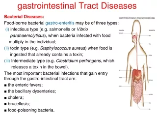
Pathological conditions of GIT • Ulcers • Zollinger-Ellison syndrome • Pernicious anemia • Malabsorption syndromes • Diarrhea
Peptic Ulcer Disease • Occurring in any part of the gastrointestinal tract exposed to the action of acidic gastric juice. • Occur principally in the duodenum (duodenal ulcer) and stomach (gastric ulcer). • Peptic ulcer occur at all ages; the most common age at onset is 20-40 years. • Duodenal ulcers are associated with blood group O, absence of blood group antigens in saliva (“non-secretors”) and the presence of HLA-B5 histocompatibility antigen.
Pathogenesis of Peptic Ulcer • Hyper-secretion of acid • - Acid is necessary for peptic ulcers to form, and ulcers do not occur in achlorhydric states. • - The cornerstone of treatment of peptic ulcer is to decrease secretion of acid; histamine H2 receptor antagonists and proton pump inhibitors are highly effective. • Decreased Mucosal Resistance to Acid • - It is believed to be the primary cause of most gastric ulcers. • - Prostaglandin E2 level in gastric juice have been shown to be consistently decreased in patients with peptic ulcer.
Helicobacter pylori Infection - In the stomach, the organism grows in the surface mucous layer, which may become altered, decreasing mucosal resistance. - H pylori can cause damage by 1) secreting urease, protease and phospholipase, 2) attracting neutrophils that release myeloperoxidase and 3) promoting thrombotic occlusion of capillaries, leading to ischemic damage of the epithelium.
Diagnosis of peptic ulcer • Based on morphological grounds (roentgenographic [photography by the use of x-ray] and endoscopic examination). • Serological tests that detect antibodies to H. pylori • Urea breath test- the individual ingests a test meal containing carbon-13 or carbon 14 labeled urea. Urease releases CO2 and the amount of labeled CO2 in breath is directly related to urease activity. • A stool antigen test for detection of H pylori.
Zollinger-Ellison syndrome • An extreme form of peptic ulcer disease, caused most commonly by a gastrin-secreting tumor of the pancreas (gastrinoma) or by antral G-cell hyperplasia of the stomach • The unrelenting gastrin release stimulates hypersecretion of HCl by the stomach • The typical clinical presentation is recurrent peptic ulceration often accompanied by diarrhea (gastrin inhibits salt and water absorption by the intestine) • The large amount of gastric interferes with fat digestion and leads to steatorrhea.
Gastric analysis • Measure secretion rate of gastric juice. • A 1 hour basal specimen is collected (basal acid output [BAO]). Reference value should be 1-6mEq/hour. • An acid production stimulant is then injected (pentagastrin) • Four 15 minutes consecutive specimens are collected. • Maximum acid output (MAO) is the sum of all four 15 min post-stimulation acid collections. Reference values for MAO are less than 40mEq/hour. • The BAO/MAO should be less than 0.3.
Diagnosis of Zollinger-Ellison Syndrome • Typically demonstrate a high basal acid secretion with minimal change after stimulation. • BAO is 15mEq/hour • BAO.MAO ratio is 0.6 or greater.
Pernicious anemia (PA) • PA is a disease that consists of gastric achlorhydria, gastric atrophy and failure to secrete intrinsic factor. • Intrinsic factor deficiency prevents absorption of Vit B12. • Vitamin B12 is an essential nutrient that is required for normal synthesis of myelin and nucleic acids. • This leads to damage to posterior columns of the spinal cord (causing a sensory neuropathy), and in many cases, megaloblastic anemia. • It is caused by autoimmune destruction of gastric mucosa (parietal cells) and intrinsic factor blocking antibodies.
Diagnosis of PA • The Shilling Test of Vit B12 absorption is used as the evaluator. • Patient should fast overnight since food may contain vit B12 and also to prevent food interference (protein bind to radioactive B12. • Oral dose of radioactive cobalt-labeled B12. Usual dose is 0.5µg. Measures the % or orally ingested isotope excreted in the urine. • 2-6 hour later, 1000µg of non-isotopic vitamin B12 is given subcutaneously or intramuscularly to saturate tissue binding sites to allow a portion of any labeled B12 absorbed from the intestine to be excreted or flushed out in the urine. • In PA, there is less than 8% urinary excretion of the radioisotope.
Malabsorption Syndromes • The syndromes are the result of any interference with the process of digestion and absorption of food. • Clinical features- loose stools, containing fat that gives a greasy appearance and foul odor to the stools (steatorrhea); loss of weight, and features secondary to fat soluble vit deficiency (bone disease, prolonged clotting times, poor night vision, neuropathy). • Can be divided into 2: true malabsorption, maldigestion. • True malabsorption- GIT is impaired, cannot absorb variety of nutrients. • Maldigestion- digestive process impaired (pancreatic insuficiency, inadequate pancreatic enzyme activation, excessive acid production, inadequate bile acid production or secretion.
Test of malabsorption • Since fat absorption is vulnerable to defects in either intraluminal digestive enzymes or defects at the mucosal absorptive surface, the demonstration of steatorrhea by qualitative or ultimately quantitative (72 hours) fecal fat measurement is the major criterion for establishing the presence of fat malabsorption.
Quantitative Fecal Fat Excretion • Anything that causes malabsorption will cause problems with fat absorption first. • Sudan staining: look for fat globules (neutral fat can be seen as bright orange droplets) • Quantitative Fecal Fat excretion: • Pt placed on 100 gram/day fat diet for 3 total days. Normal excrete ~ 3-5 grams fat/day. > 15 grams excretion over the 3 days is (+) result 2 SD > normal mean. • Can easily get false positive/negative: Poor food intake, constipation, forgot to collect stool. Eating nuts that cause fat in stool.
D-Xylose Absorption-Excretion Test • Dz that involves epithelium or mucosa itself. • D-xylose is a water soluble, non-metabolized pentose that is absorbed mainly in the duodenum and jejunum. No intraluminal handling. No pancreatic enzymes needed. Will normally absorb across epithelium. Assimilation of this sugar does not require the intraluminal pancreatic stage of digestion.
D-Xylose Absorption-Excretion Test Test procedure • The standard test dose is 25gm of D-xylose in 250 ml of water, followed by another 250ml water. • The pt is fasted overnight, since xylose absorption is delayed by food. • The normal person’s peak D-xylose are reached in approximately 2 hours and fall to fasting levels in approximately 5 hours. • D-xylose 80-95% is excreted mostly in the urine in the first 5 hour and the remainder 24 hour later.
Interpretation • Normal values for 2 hour blood D-xylose levels are more than 25mg/100ml. • Values less than 20mg/100ml are strongly suggestive of malabsorption. -Abnormal test: Intestinal mucosal disease. Also with the small intestinal bacterial overgrowth. · Normal test: deficiency of intraluminal (pancreatic) digestive enzymes or bile acid deficiency
PABA Test to Evaluate Pancreatic function • Test is done w/PABA (ρ-aminobenzoic acid) and an attached tripeptide. If have exocrine function then will cleave off tripeptide. • Normal function: Ingest PABA-tripeptide tripeptides cleaved liberating PABA which is absorbed excreted in urine. • Abnormal: no cleavage of tripeptide. Is absorbed, but PABA is not excreted in urine.
Other Tests for Malabsorption • Pancreatic Function Tests. The secretin test is used to measure secretory capacity of the exocrine pancreas. After administration of secretin, bicarbonate concentration is measured in the juice aspirated from the duodenum. • Measurements of serum iron, calcium, cholesterol, folate and vitamin B12 often are used as screening tests for malabsorption, but are not specific. • Prothrombin Time. If prolonged, may reflect malabsorption or liver disease. These possibilities can be distinguished by measuring the response to parenterally administered vitamin K. • Serum Carotene. Carotene is a fat-soluble substance present in yellow vegetables and fruits, eggs, etc. Serum carotene levels tend to be depressed in patients with fat malabsorption, but can be decreased also if the intake of dietary carotene is low.
Diarrhea • Excessive production of feces, usually as a result of overabundance of water in the stool. • Severe diarrhea causes sodium and water depletion and loss of potassium and bicarbonate. • There are 2 causes of diarrhea: 1) decreased absorption of fluid and electrolytes. 2) increased secretion of fluid
Decreased Absorption of Fluid and Electrolytes • Decreased absorption of fluid and electrolytes in intestinal malabsorption. • Absorption of water in the intestines is dependent on adequate absorption of solutes. If excessive amounts of solutes are retained in the intestinal lumen, water will not be absorbed and diarrhea will result (osmotic diarrhea). • Ingestion of a poorly absorbed substrate: The offending molecule is usually a carbohydrate or divalent ion. Common examples include mannitol or sorbitol, epson salt (MgSO4) and some antacids (MgOH2). • Malabsorption:lactose intolerance resulting from a deficiency in the brush border enzyme lactase.Lactose cannot be effectively hydrolyzed into glucose and galactose for absorption. The osmotically-active lactose is retained in the intestinal lumen, where it "holds" water.
Increased secretion of fluid • Vibrio cholerae, produces cholera toxin, which activates adenylyl cyclase, causing a prolonged increase in intracellular concentration of cyclic AMP within crypt enterocytes. This change results in prolonged opening of the chloride channels that are instrumental in secretion of water from the crypts, allowing uncontrolled secretion of water. Additionally, cholera toxin affects the enteric nervous system, resulting in an independent stimulus of secretion. • In addition to bacterial toxins, a large number of other agents can induce secretory diarrhea by turning on the intestinal secretory machinery, including: • some laxatives (foods, compounds, or drugs taken to induce bowel movements or to loosen the stool, most often taken to treat constipation). • hormones secreted by certain types of tumors (e.g. vasoactive intestinal peptide) • a broad range of drugs (e.g. some types of asthma medications, antidepressants, cardiac drugs) • certain metals, organic toxins, and plant products (e.g. arsenic, insecticides, mushroom toxins, caffeine)
Diarrhea Associated with Deranged Motility • In order for nutrients and water to be efficiently absorbed, the intestinal contents must be adequately exposed to the mucosal epithelium and retained long enough to allow absorption. • Disorders in motility than accelerate transit time could decrease absorption, resulting in diarrhea