Download
1 / 20
230 likes | 496 Vues
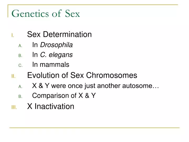
Genetics of Sex. Sex Determination In Drosophila In C. elegans In mammals Evolution of Sex Chromosomes X & Y were once just another autosome… Comparison of X & Y X Inactivation. I. Sex determination mechanisms.

E N D
Genetics of Sex • Sex Determination • In Drosophila • In C. elegans • In mammals • Evolution of Sex Chromosomes • X & Y were once just another autosome… • Comparison of X & Y • X Inactivation
I. Sex determination mechanisms • Although most animals have two sexes (M/F) there is a great variety of mechanisms that have evolved • GSD • X:A • XX/XY • ZW/ZZ • ESD
Sex Determination: A. in Drosophila • Every cell determines its sex independently • Ratio of X chromosomes to Autosomes is what determines sex
Early in embryonic development, a cascade of differential (alternate) mRNA splicing results in either female or male. • The three branches of the hierarchy govern: • X chromosome dosage compensation • somatic sexual differentiation • male sexual behavior
= 1 = 0.5
B. C. elegans – hermaphrodites or males? • Like Drosophila,
X:A = 1, enhanced expression of fox-1 & sex-1whose products inhibits the expression of xol-1. sdc genes are expressed, which are involved in dosage compensation and hermaphodite development.
C. Sex determination in mammals • Not independent for each cell • Not as simple a pathway as Drosophila or C. elegans… not yet completely understood • Sex is determined by the presence or absence of the Y chromosome
Gonad is bipotential • 3 cell lineages present in gonad as well as the germ cells • Supporting cell lineage will give rise to Sertoli cells in testis & follicle cells in ovary • Steroidogenic cell lineage – produce sexual hormones • Each lineage has
Mammalian gonad forms within the developing urogenital system, which itself derives from the intermediate mesoderm. This is divided into 3 regions: Epithelial structures are shown in red, mesenchymal structures are shown in blue, and the striped region denotes the genital ridge. (WD) Wolffian duct; (MT) mesonephric tubules; (MD) Mullerian duct; (UB) ureteric bud; (CE) coelomic epithelia.
II. Evolution of the X & Y A. X & Y were once just another autosome… • Evolved 300 mya, autosomal origin • X – 1,098 genes (lowest # compared to autosomes) • 4,493 ECRs conserved between human, mouse, rat, zebrafish & pufferfish • Males Hemizygous – • X linked inheritance
Autosomal origin of X supported by orthologous regions from Chicken, 30 regions of homology illustrated:
B. Comparison of X & Y • Y significantly smaller than X, few genes shared between the two • In the Y chromosome, the shutting down of X–Y crossing over during evolution triggered a monotonic decline in gene function • PAR1 homology maintained by recombination in male meiosis, genes in this region not subject to dosage compensation
III. X Inactivation • Dosage compensation mechanism in mammals • Mammalian cells have ability to count their X chromosomes • X inactivation center (Xic) plays critical role • Xist (located in Xic) expression required for compaction of X into a Barr body • X chromosomal controlling element (Xce) & TsiX affects the choice of which X to be inactivated • About 15% of the genes escape effects of X inactivation • Xist • Pseudoautosomal genes also found on Y • Female sex-specific genes http://www.hhmi.org/biointeractive/animations/x_inactivation/xinact_frames.htm