DENTAL RADIOGRAPH.
DENTAL RADIOGRAPH. SHAMA ISMAIL 12BME28. Introduction . Dental X-ray machine also called Dental Radiograph, are the most valuable tools, a dentist has for keeping your mouth and teeth health.

DENTAL RADIOGRAPH.
E N D
Presentation Transcript
DENTAL RADIOGRAPH. SHAMA ISMAIL 12BME28.
Introduction • Dental X-ray machine also called Dental Radiograph, are the most valuable tools, a dentist has for keeping your mouth and teeth health. • Dentists use radiographs for many reasons: to find hidden dental structures, malignant or benign masses, bone loss, and cavities • Alert the dentist to possible bone loss associated with periodontal disease
Continued…. • Reveal problems in the root canal, such as infection or death of the nerve. • Reveal other abnormalities such as cysts, cancer. • For children, radiographs are used to watch for decay and to monitor tooth growth and development
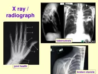
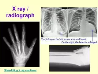
Types of X-rays • X-rays are divided into two main categories: • Intraoral: which means that the X-ray film is inside the mouth; • Extraoral: which means that the film is outside the mouth
Intraoral X-rays • Are the most common type of X-ray taken. • These X-rays provide a lot of detail and allow your dentist to find dental caries. • Check the health of the tooth root and bone surrounding the tooth, • Check the status of developing teeth, and monitor the general health of your teeth and jawbone.
Different Types of Intraoral X-ray • Bite-wing Dental X-rays. • Periapical Dental X-rays. • Occlusal Dental X-rays
Bite-wing Dental X-rays • Bite-wing X-rays show details of the upper and lower teeth in one area of the mouth. • Each bite-wing shows a tooth from its crown to about the level of the supporting bone. • Bite-wing X-rays are used to detect decay between teeth and changes in bone density caused by gum disease. • They are also useful in determining the proper fit of a crown
Continued…. Bitewing dental X-ray unit. Bitewing dental X-ray
Periapical Dental X-rays • The name periapical is derived from the Greek language : peri, which means "around," and apical, which means "tip. • Periapical X-rays are used to detect any abnormalities of the root structure and surrounding bone structure. • it highlight the entire tooth
Occlusal Dental X-rays • Occlusal X-rays are larger and highlight tooth development and placement. • On each radiograph, nearly the full arch of teeth in either the upper or lower jaw is shown. • These X-rays are taken with the X-ray machine either pointing straight down from near the nose to take pictures of the upper jaw and teeth, or straight up from the chin to take picture of the lower jaw and teeth.
Extraoral Dental X-Ray • Extraoral X-rays are made with the film outside the mouth. • These can be considered the " big picture" X-rays. • They show teeth, but their main focus is on the jaw or skull. • Extraoral X-rays are less detailed than intraoral X-rays, so they are not used for detecting caries or flaws in individual teeth.
Continued…. • Extraoral radiographs are used for monitoring growth and development, looking at the status of impacted teeth, examining the relationships between teeth and jaws and examining the temporomandibular joint or other bones of the face.
PANORAMIC RADIOGRAPH. • Panoramic X-rays show a broad view of the jaws, teeth, sinuses, nasal area, and temporomandibular (jaw) joints. • These X-rays do not find cavities. • One advantage of the panoramic X-ray is its ease of use. Unlike other X-rays where the film is placed inside the patient's mouth, the panoramic film is contained in a machine that moves around the patient's head
Continued…. • These X-rays do show problems such as impacted teeth, bone abnormalities, cysts, solid growths (tumors), infections, and fractures. • Panoramic radiograph takes a picture of all the bony structure of the face.
How should patient prepare? • A panoramic x-ray examination requires no special preparation. • You will be asked to wear a lead apron as a safety precaution to protect the rest of your body from any radiation exposure that may scatter from the panoramic x-ray beam. You may also be asked to remove your jewelry from the region being imaged, eye glasses and any metal objects that might interfere with the x-ray images.
Continued…. • Women should always inform their dentist or oral surgeon if there is any possibility that they are pregnant. • Many imaging tests are not performed during pregnancy so as not to expose the fetus to radiation. • If an x-ray is necessary, precautions will be taken to minimize radiation exposure to the baby.
What does the equipment look like? • A panoramic x-ray machine consists of an x-ray tube mounted on a horizontal arm. • X-ray film are mounted on another horizontal arm on the opposite side of the patient. • For a sharper, clearer image, the patient's head is positioned with chin, forehead and side rests. • The patient is also provided with a bite blocker to properly open the oral cavity.
How does the procedure work? • X-rays are a form of radiation like light or radio waves. X-rays pass through most objects, including the body. Once it is carefully aimed at the part of the body being examined, an x-ray machine produces a small burst of radiation that passes through the body, recording an image on photographic film or a special detector.
Continued…. • During a panoramic x-ray examination, the x-ray tube rotates in a semicircle around the patient's head, starting at one side of the jaw and ending at the other side. • Rather than relying on film placed inside the mouth, a panoramic x-ray machine projects a beam through the patient onto film or a detector rotating opposite the x-ray tube. • x-ray images were maintained as hard film copy
Continued…. • Today, most images are digital files that are stored electronically. These stored images are easily accessible and are frequently compared to current x-ray images for diagnosis and disease management. • The digital format also allows the dentist to adjust and change the contrast, brightness and darkness of the image for better visualization of certain structures and tissues.
Continued…. • Images on film cannot be adjusted or changed
How is the procedure performed? • This examination is usually done on an outpatient basis. • First, you will be situated in the center of the unit where the technician will carefully position and secure your head. The unit can be adjusted to accommodate a patient standing or sitting in a wheelchair.
Continued…. • A bite-blocker is then placed in your mouth to ensure proper alignment of the teeth. Correct placement of the teeth and head is important for obtaining a clear image.
Continued…. • You will be asked to remain very still while the rotating arm travels in a semicircle around the perimeter of your head and the images are being recorded, which can typically take between 12 to 20 seconds. • Any movement from the patient will blur the image on the screen.
Dental X-ray Machine • The most common used Dental X ray machine is the wall-mounted dental x-ray unit. • Because of the basic components and operating techniques of all dental x-ray machines are similar, that’s why we only discuss the wall-mounted unit.
PARTS AND COMPONENTS OF THE DENTAL X-RAY MACHINE • Tube Head. • Cylinder. • The Extension Arm. • The Control Panel.
Tube Head. • Inside the metal tube housing is the x-ray tube. The diagram in next figure represents a dental x-ray tube head. • This tube emits radiation in the form of photons. X-ray photons expose the film. In addition to exposing the film, it also exposes the patient to radiation. Unless certain protective measures are taken, the x-ray technician may also be exposed.
Cylinder. • The cylinder (cone) is affixed to the tube head and is used to align the tube head with the patient and x-ray film.
Extension Arm. • The tube head is attached to the metal extension arm by means of a yoke that can revolve 360 degrees horizontally where it is connected. The construction of the yoke also provides vertical movement as well
Control Panel. • The components of the control panel are switches, dials, gauges, and lights. • Energize the control panel. • Set the tube head selector. • Select the kilo voltage. • Set the exposure time. • Make the exposure by depressing the exposure button located on the control panel. • After making the exposure, process the X-ray film.