Download
1 / 19
190 likes | 210 Vues
Active Pixel Sensors in Medical and Biologi. The application of Large Area Active Pixel Sensor (LAS) to high resolution Nuclear Medicine imaging Bob Ott Physics Department Institute of Cancer Research and Royal Marsden Hospital. What is an active pixel sensor (APS).

E N D
Active Pixel Sensors in Medical and Biologi The application of Large Area Active Pixel Sensor (LAS) to high resolution Nuclear Medicine imaging Bob Ott Physics Department Institute of Cancer Research and Royal Marsden Hospital
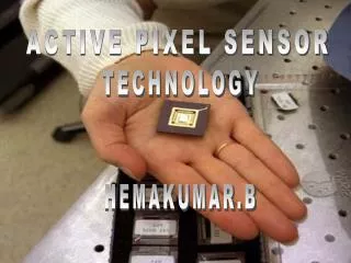
What is an active pixel sensor (APS) • A silicon wafer based sensor similar to a CCD but with the potential for intelligent processing in each pixel • APSs are based on mainstream CMOS technology and have generic benefits of low-power, high-speed, cost-effectiveness, flexibility and high levels of on-chip integration.
Other APS properties Back thinning allow low energy beta/gamma/X-ray radiation to be detected Devices can be used either as direct sensors or coupled to phosphors for example. Devices can be read out through special FPGA based interfaces to allow data acquisition onto a PC or laptop. Potential for radiation hardness Potential for ROI read-out
The MI-3 research consortium MI-3 stands for Multidimensional Integrated Intelligent Imaging MI-3 was a four-year £4.5m Basic Technology project funded by UK-RC to advance the capabilities and application of APSs for a raft of scientific and technological endeavours Consortium members Universities of Sheffield, Liverpool (2 groups), Glasgow, Surrey, York, Brunel, UCL and Rutherford Appleton Laboratories MRC Laboratory for Molecular Biology, Institute of Cancer Research.
MI-3 APS applications Medical and biological imaging (UCL, Surrey, ICR) Space science (Brunel) Bioarray imaging (Liverpool) Electron microscopy (Cambridge) Particle physics (Liverpool, Glasgow) Synchrotron radiation applications (Sheffield) Materials science (York) Detector Design and testing mostly done at RAL/STFC
Properties of APSs used in this project *Indicate the noise range which is dependent on the reset type. **The rates depend on whether full-frame or ROI read-out is used.
High-resolution Nuclear Medicine imaging requirements Pixel sizes of 50-100 microns Coupling to CsI(Tl) phosphors to detect 140keV photons Low noise needed as signal is small >100 cm2 sensitive area Kcps photon counting capacity On-chip pulse and cluster analysis for signal selection and noise reduction
High-resolution gamma camera APS coupled to pixelated CsI(Tl) phosphor via optical fibre stud
High-resolution gamma camera 5mm wide slot System resolution is ~1mm FWHM Using 2mm thick CsI(Tl) scintillator with 400µm elements coupled to Vanilla
LAS Pixel size 40 µm x 40 µm 1350 x 1350 pixels 54 mm x 54mm stitched sensor Read-out noise between 40e and 62e 73% fill factor Region of interest read-out available Maximum frame rate 20s-1 QE at 520 nm ~22% (Vanilla >60%) Average QE between 500nm and 800nm ~16% Bohndiek et al, IEEE TNS, 56, 2938-2946, 2009
Dark image taken with LAS Horizontal profile
X-ray resolution phantom imaging 0.1 lp mm-1 0.25 lp mm-1 Taken with a 99mTc flood source
LAS coupled to unsegmented 2mm thick CSI(Tl) flood source FWHM of ~ 2.5mm
LAS flood image of 20mm x 20mm segmented CsI(Tl) scintillators 400µm elements 350µm elements
Problems and solutions • The signal from Tc-99m is small so that segmented CsI (Tl) is important • Higher QE (from <20% to >60%) essential • Processing to remove fixed pattern noise improves image quality • Larger CMOS pixels with low noise preferred • Larger area than LAS needed – see Dynamite
Dynamite – coming soon Active area 120 mm x 120 mm Two pixel sizes – 50 µm and 100 µm 15 frames/s for full read-out Faster for ROI read-out Pixelated CsI(Tl) scintillator available
Conclusions APSs could make a contribution to Nuclear Medicine imaging Noise level requirements are similar to CCDs but with faster readout speed Fixed pattern noise dominates over thermal noise On-chip intelligence for noise filtering and allow ROI selection Large area devices essential for this application Dynamite will be very interesting