Download
1 / 51
520 likes | 552 Vues
This session explores the significance of respiratory assessment in patient care, covering initial quick assessment, patient monitoring techniques like pulse oximetry and capnography, as well as components like history and physical examination. Learn to identify respiratory issues, administer proper treatment, and evaluate response for effective patient management. Enhance your skills in recognizing abnormal breath sounds, interpreting chest X-rays, and monitoring patient responses. Join us for an informative session on optimizing respiratory assessment practices.

E N D
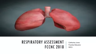
Respiratory assessmentFCCNC 2018 Catherine Jones Practice Educator GICU
Plan for session • 1. Why bother with Respiratory Assessment? • 2. Patient Assessment • History • Look, Listen, Feel • 3. Patient Monitoring • Pulse Oximetry • Capnography • Vent Graphics
Why do we assess? • To ensure safety • To have a baseline that we can build our goals and management (identify issues) • To prevent or identify problems • Respiratory system is responsible for external and internal respiration (gas exchange) • To administer most appropriate treatment • To evaluate treatment’s response
Components of respiratory assessment 1. Initial quick assessment 2. History • PMH, medication, allergies 3. Patient Assessment • Look • Listen • Feel 4. Patient Monitoring • Pulse Oximetry (SpO2) • End Tidal Co2 Monitoring (EtCO2) • Ventilator Graphics • Arterial blood gas analysis (session tomorrow) • Chest XRay 5. Documentation
1. Initial Assessment AKA ‘End of bed ogram..’ • Patient Responsive or Unresponsive? • Unresponsive? BLS
3. Patient assessment FEEL LISTEN LOOK
LOOK Colour Position Chest Shape & Scars Ability to speak Respiratory Rate, depth & Pattern Accessory Muscles Chest Movement Sputum – quantity, viscosity, colour
Normal Adults:12-20/min Tachypnea Rapid, shallow breathing Hyperypnea Rapid, deep breathing Hyperventilation Kussmaul breathing Bradypnea Breathing Patterns • Rate, Depth, Regularity Ataxic breathing Biot’s breathing Irregularly irregular Cheyne-Stokes breathing Regular rate, irregular depth MAY be normal Sighs Hyperventilation syndrome 1 sigh per 200 breaths
21 secs clipflail chest & paradoxical breathing https://www.youtube.com/watch?v=mJ_FYwUqzsM Usually associated with chest trauma flails & #ribs or extreme fatigue 2ndry acute respiratory failure
LISTEN • Speech • Noisy respirations - Stridor, wheeze, rattly chest, gurgling, snoring
Stridor • High pitched wheezing sound caused by obstruction to airflow. • Usually blockage to larynx, trachea (anaphylaxis, tumour, swelling..) • BAD SIGN – If new onset – GET HELP!!
Chest Auscultation • Movement of air in & out of airways • Flow and airway size dependent • Normal sounds / Added abnormal sounds • Interpretation Practice!
CLASSIFICATION OF BREATH SOUNDS: normal • Vesicular • Quiet breath sounds heard over most lung fields. • Bronchial • Loud, hollow, high pitched breath sounds heard over bronchus. • NB this is an ABNORMAL sound if heard elsewhere – indicative of consolidation. • Bronchiovesicular • Normal breath sounds heard over lung tissue between bronchus & peripheries
CLASSIFICATION OF BREATH SOUNDS: Abnormal • Diminished / Absent? • Obesity • tidal volumes • Pleural effusion • Occluded airway + collapse • Pneumothorax
CLASSIFICATION OF BREATH SOUNDS: Abnormal • Added sounds • Wheeze • Crackles • Stridor (NB Don’t need auscultation to hear) • Pleural Rub www.wilkes.med.ucla.edu/intro.html: normal; wheeze & crackles
CHEST X-RAY - NORMAL • Name & date • Technical quality Exposure AP / PA Orientation / Rotation Degree of inspiration • Methodically review film
FEEL Tracheal Deviation Surgical emphysema - presence of air in s/c tissue Equality of chest expansion/excursion Tactile fremitus Technique – Pt says ‘99’ resulting in a palpable vibration Decreased transmission = pleural effusion, pneumothorax Increased transmission = pneumonia, consolidation MV – fremitus can be found when there are secretions in airways
4. Patient monitoring PULSE OXIMETRY VENTILATOR WAVEFORMS CAPNOGRAPHY
PULSE OXIMETRY • SpO2 is an indirect measurement of peripheral arterial oxygen saturation • Red & Infrared Light are emitted from a diode through pulsatile blood flow to a photodector. • Oxygenated (Bright Red) blood differs from deoxygenated (Blue Tinge) blood in light absorption & the oximeter measures the amount of light absorbed over 5 pulses to calculate the oxygen saturation.
Limitations of pulse oximetry • Doesn’t give any info about adequacy of ventilation • Dependent on adequate peripheral perfusion • Cardiac Arythmias • Motion artefact – pt movement or shivering • Nail varnish – equivocal evidence – blue, black, green • Dyshaemoglobins – carboxyhaemoglobin & methaemoglobin – can’t distinguish from oxyhaemoglobin so false high. • Skin pigment - jaundice
Capnography MAINSTREAM SAMPLING SIDE STREAM SAMPLING
Why Measure ETCO2? • Monitor adequacy of ventilation • Detect correct placement of ETT • Detect apnoea • Indication of acid base balance • Indication of cardiac output • Indication of metabolic state • Monitor pulmonary perfusion in CPR • Diagnosis of PE • Safety in transporting critical care patient
Measurement of CO2 at the very end of expiration • Correlates with PaCO2 in ‘normal lungs’ <1kpa difference • EtCO2 can be significantly lower than PaCO2 (with poor lungs or poor perfusion)
General info • Ventilator Waveforms depend on • Mode of ventilation • Ventilator properties & settings • Patient lung characteristics • 2 Types of Waveform: • Scalar (aka Curve) Pressure/Time; Volume/Time; Flow/Time • Loops Pressure/Volume; Volume/Flow; Flow/Pressure
Final thoughts • Respiratory data can be sought from multiple sources • ‘Old fashioned’ hands on assessment complements ‘New fashioned’ hands off assessment
references • Coombs et al (2013) ‘Assessment, monitoring & interventions for the respiratory system’, in Mallet et al (Eds) Critical Care Manual of Clinical Procedures & Competencies (Wiley Blackwell, London) Pgs. 63-166) • Corley A. & Ringdal M. (2012) ‘Respiratory Assessment & Monitoring’, in Elliott et al (Eds) ACCCN’s Critical Care Nursing 2nd ed (Mosby Elsevier, London) Pgs 325-351) • Moore T. Respiratory Assessment in Moore T. & Woodrow P. (2004) High Dependency Nursing Care (Routledge, London) P.124-133. • Smith J. & Rushton M. (2015) How to perform respiratory assessment Nursing Standard 30,7. P.34-36.