Download
1 / 19
210 likes | 1.2k Vues
Assessment of the respiratory system. Prepare by: Mrs. Mahdia Samaha Alkony. 1. Health History. The reason for seeking advice: dyspnea, hemoptysis, odema, cough, general fatigue, weakness. Chief complain , onset , duration, severity Assess risk factors

E N D
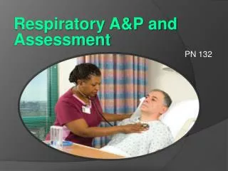
Assessment of the respiratory system Prepare by: Mrs. Mahdia Samaha Alkony 1
Health History • The reason for seeking advice: dyspnea, hemoptysis, odema, cough, general fatigue, weakness. • Chief complain , onset , duration, severity • Assess risk factors • Identify the impact of signs& symptoms on the patient ability to perform daily activities. 2
Major signs and symptoms of the respiratory diseases: • Dyspnea: difficulty in breathing, shortness of breath. Symptom common when there is decrease lung compliance or increase airway resistance. Sudden dyspnea in healthy person indicates: • Pneumothorax. • Pulmonary embolism • RDS. • Acute respiratory obstruction.
Major signs and symptoms of the respiratory diseases: 2.Orthopnea: inability to breath easily except in an upright position. Found in pt. with COPD, heart disease. • Noisy breathing result from narrowing of the airway or localized obstruction of major bronchus by tumor or foreign body. 3.Cough: caused byirritation of the mucous membranes of the respiratory tract that may arise from an infectious process, irritant as smoke, dust… It’s a protective mechanism against accumulation of secretion in the bronchi & bronchioles.
Charachters of cough • A dry, irritant cough is an URTI of viral origin • High pitched cough laryngotracheitis caused by an irritation. • A cough in the morning with sputum production may indicate bronchitis. • A cough that worsen when the pt. is in supine position suggest postnasal drip (sinusitis). • Coughing after food intake may indicate aspiration of material into the tracheobronchial tree.
Major signs and symptoms of the respiratory diseases: 4- Sputum production: • Thick, yellow, green; Indicate bacterial infection. • Thin, mucoid sputum; Indicate viral infection. • Pink tinged mucoid sputum; Indicate lung tumor. • Profuse, frothy, pink material ; Indicate pulmonary odema. • Foul-smelling sputum & bad breath ; Indicate lung abscess, infection from anaerobic organism.
Major signs and symptoms of the respiratory diseases: • Cough management: • Adequate hydration (water). • Inhalation of nebulizer. • Stop smoking.
5-Chest pain: Chest pain may be sharp stabbing & intermittent or dull, aching & persistant: Causes of chest pain: • It is a late symptom of bronchogenic carcinoma. • Pneumonia, pulmonary embolism, lung infarction & pleurisity. • Pleuritic pain from irritation of partial pleura is sharp (like the stabbing of knife). Pt. becomes comfortable when sleeps on the affected side. • Pain must assessed for quality, intensity, radiation of pain, relationship of pain to inspiratory & expiratory.
Nursing interventions for chest pain: • Analgesic medication but not to depress the respiratory center or productive cough. • NSAID for pleurituic pain. • Regional anesthetic block may be performed to decrease extreme pain.
Major signs and symptoms of the respiratory diseases: 6- Wheezing: Is a major finding in pt. with bronchoconstriction or airway narrowing. Wheezing is a high pitched, musical sound heard mainly on expiration. Relived by: Oral or inhaled bronchodilator. 7- Clubbing of the fingers: is a sign of chronic hypoxic condition, chronic lung infections, CA of lung.
Major signs and symptoms of the respiratory diseases: 7- Hemoptysis: Is symptom of both pulmonary & cardiac disorders. • Onset is sudden, may be intermittent or continuous. Common cause: • Pulmonary infection. • CA of lung. • Abnormalities of heart & blood vessels. • Pulmonary artery or vein abnormalities. • Pulmonary emboli & infarction.
Major signs and symptoms of the respiratory diseases: 9- Cyanosis: Is a bluish coloring of skin, very late indicator of hypoxia. • Cyanosis is not reliable singe of hypoxia. Because anemic pt. rarely manifest cyanosis. • In the presence of pulmonary disease cyanosis assessed by tongue & lips. • Peripheral cyanosis results from decrease blood flow to certain area & not indicate central problem.
Diagnostic evaluation • Pulmonary function test. Routinely used in pt. with chronic respiratory disorders, test should measurements of lung volume, ventilatory function & mechanism of breathing diffusion & gas exchange. • Arterial blood gas study. Aid in assessing the ability of the lungs to provide adequate O2 & remove CO2, the ability of kidney to reabsorb & excrete bicarbonate ions to maintain body normal PH.
Diagnostic evaluation • Pulse oximetry; Monitor O2 sat. of hemoglin. Normal O2 sat. is 95-100%, decrease 85 indicate that tissue are not receiving enough o2. • Culture; Specimen to lab must be within 2 hr.s (overgrowth of organisim). Specimen taken at morning. • Imagining studies;. 1-Chest x-ray: normal pulmonary tissue is radiolucent, there for densties produced by fluid, tumors, foreign bodies & pathogenic condition can be detected by x-ray. 2-CT : used to identify pulmonary nodules & small tumors that are not visible on routine chest x-ray.
Diagnostic evaluation 3-MRI :are more diagnostic image than CT to characterized pulmonary nodules. 4-Fluoroscopic studies: used to detect diaphragm paralysis & lung masses. 5-Pulmonary angiography: used to investigate thromboembolic disease of the lung as congenital abnormalities of pulmonary vascular tree. 6-Radioisotope (lung scan).Ventilation- perfusion lung scan to measure the integrity of pulmonary vessels to evaluate blood flow abnormalities as seen in pulmonary emboli.
VI. Endoscopic procedure; Bronchoscopy is direct inspection & examination of the larynx, trachea, & bronchi through either a flexible fiberoptic scope or rigid bronchoscope. Diagnostic Bronchoscopy. • To examine tissue or collect secreations. • To determine location & obtain tissue. • To determine if tumor can be resected surgically. • To DX. Bleeding site. Therapeutic bronchoscopy : • Remove foreign bodies. • Remove Secreation from the tracheobronchial tree. • Treat post operative atelactasis. • Destroy & excise lesion.
NSG intervention: • consent form • Keep NPO • Explain procedure • Preoperative medication ( atropine, sedation) that inhibit vagal stimulation, suppress cough. • Post op. pt. must be NPO until the cough reflex returns. • Ice-chips & fluid given. • Observe v/s, hypoxia, bleeding, hypotention, tachycardia, dysarythmia.
Thoracentesis • Aspiration of fluid from the pleural cavity for DX. Or therapeutic purposes). • Study includes grams stain culture & sensitivity, acid- fast staining, PH.