Understanding Amino Acids: Structure, Abbreviations, and Techniques for Analysis
240 likes | 356 Vues
This article delves into the structure and abbreviations of amino acids, highlighting their crucial roles in biochemistry. It explains the significance of 1-letter and 3-letter abbreviations, such as Gly for Glycine and Ala for Alanine. The concept of isoelectric points (pI) is introduced, detailing how they influence amino acid charge and separation techniques like ion exchange chromatography and polyacrylamide gel electrophoresis. Furthermore, methods for determining amino acid sequences using selective hydrolysis and derivatization are explored, emphasizing analytical techniques for peptide analysis.

Understanding Amino Acids: Structure, Abbreviations, and Techniques for Analysis
E N D
Presentation Transcript
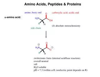
Amino Acids: Structure, Analysis, and Sequence (in peptides)
Abbreviations of Amino Acids • Amino acids have 1-letter and 3-letter abbreviations; the 1-letter abbreviations are used almost exclusively today, but you should also be aware of the older 3-letter abbreviations. • Some examples: • glycine (R = H) Gly G • alanine (R = CH3) Ala A • phenylalanine (R = CH2C6H5) Phe F • tyrosine (R = CH2C6H4OH) Tyr Y • serine (R = CH2OH) Ser S • cysteine (R = CH2SH) Cys C • methionine (R = CH2CH2SCH3) Met M • leucine (R = CH2CH(CH3)2) Leu L
Isoelectric Point • Each amino acid has an isoelectric point, (pI) numerically equal to the pH at which the zwitterion concentration is at a maximum. • The amino acid has no NET charge at its pI; it has one positive and one negative charge. • At a pH less than the value of the isoelectric point, the amino acid is protonated and has a POSITIVE charge; at a pH greater than the pI the amino acid is deprotonated and has a NEGATIVE charge. Cation Neutral Anion (zwitterion form)
Separation and Analysis using pI values • Differences in isoelectric points (and therefore charges) are used to separate mixtures of amino acids by two common methods: • Ion exchange chromatography • Polyacrylamide gel electrophoresis (PAGE) These methods will be illustrated with a simple mixture of three amino acids having very different isoelectric points: aspartic acid alanine lysine
Ion Exchange Chromatography D- elutes first, followed by A; K+ elutes last, and only after pH of buffer is increased and K+ is deprotonated.
Ion Exchange Chromatography • Recall that in our simple mixture D- elutes first, followed by A; K+ elutes last, and only after the pH of buffer is increased and K+ is deprotonated. • But there is a problem in detecting amino acids; they are colorless, and most of them have very little absorption in the UV region (they have no conjugation, except in the four aromatic amino acids) • To overcome this difficulty, amino acids are converted (after separation by ion exchange chromatography) to a derivative using ninhydrin.
Derivatization with Ninhydrin Ninhydrin (2 mol) reacts with one mol of ANY amino acid to give the SAMEblue colored product. This reaction is performed post-column, after Ion Exchange Chromatography separation of a mixture of amino acids. The area of each peak in the chromatogram is proportional to the relative molar amount of the amino acid of that retention time.
Ion Exchange Chromatography Recall that in our simple mixture D- elutes first, followed by A; K elutes last, and only after the pH of buffer is increased and K+ is deprotonated. injection Increase pH of buffer Retention time
Polyacrylamide Gel Electrophoresis (PAGE) Before current is turned on: After current is turned on:
The Strecker amino acid synthesis (racemic alanine)
Resolution of racemic amino acids D- L- L-amino acid + D-N-acetylamino acid Racemic amino acid Racemic N-acetyl amino acid Carboxypeptidase hydrolyzes the amide bond ONLY of the L-aa, leaving the unnatural D-N-acetylamino acid unreacted; separation is simple
Covalent bonding in peptides • Amino acids are covalently bonded to one another by amide linkages (bonds) between the carboxylic acid group of one amino acid and the amino group of the next amino acid. • Amide bonds are strong and are resistant to hydrolysis, but there are enzymes that catalyze their hydrolysis (to the amino acids). • In addition to amide bonds, a second kind of covalent bond exists in some peptides in which two cysteine residues (amino acid units) are connected through a disulfide bond formed by oxidation (dehydrogenation) of the sulfhydryl (SH) groups (next slide).
Total Hydrolysis: conversion of a peptide into a mixture of its component amino acids Ion Exchange Chromatogram:
2. Amino Acid Sequence: Primary Structure Determination of Peptides • Total hydrolysis followed by ninhydrin derivatization and ion exchange chromatography tells us the identity and relative amount of each amino acid present in the peptide • It gives NO INFORMATION about the sequence, or order of attachment of the amino acids, however. • For this, we need to perform selective hydrolysis of the peptide. • We’ll learn three methods: • Sanger’s reagent followed by total hydrolysis • Carboxypeptidase • Leucine aminopeptidase
Partial Hydrolysis Peptide represented schematically: (some molecules) (other molecules) (some other molecules) (different molecules of the peptide can fragment differently, leading to a mixture)
Putting it all together! • Suppose an unknown hexapeptide gave “tagged” A (alanine) upon treatment with Sanger’s reagent, and upon treatment with carboxypeptidase, the first amino acid released was M (methionine) followed by G (glycine) • Partial hydrolysis gave the following identifiable tripeptides: V-G-M, A-S-F, and S-F-V. What is the 1º structure of the hexapeptide? A S F V G M A S F V G M V GM AS F V GM AS F V GM A S F V GM