Download
1 / 60
610 likes | 771 Vues
Amino acids are the essential building blocks of peptides and proteins, playing pivotal roles in biological processes. They exist in various forms, including essential amino acids that must be obtained from diet. This overview covers amino acid structures, functionalities, and classifications, including zwitterions and amphoteric properties. It explains polypeptide formation through dehydration reactions, highlighting significant structural levels (primary, secondary, tertiary, and quaternary) and the importance of hydrogen bonds, ionic interactions, and disulfide bonds in protein functions like catalysis, transport, and structural support.

E N D
Amino Acids General
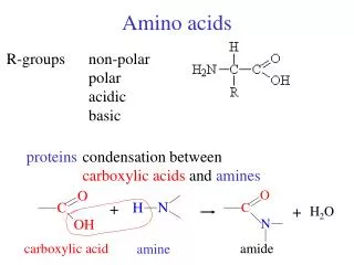
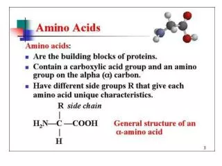
Amino Acids: • Building blocks for peptides, proteins • Some individually important (or converted to important molecules) • Gly, Glu, Tyr neurotransmitters • Tyr parent/precursor for epinephrine (adrenaline) • His stomach secretes HCl, symptoms for inflammation, colds. • Essential (10) • needed for normal health • not synthesized by the body • must be supplied by diet • Complete (animal) vs. Incomplete (vegetable) protein
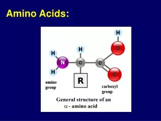
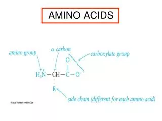
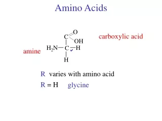
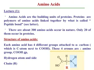
Amino Acids Structure
Amino Acid Structure: • Amide, CA, R-group (variable) • D/L Isomers
Amino Acids Side Chains
AA – Side Chains: • Side chains determine the functionality of the AA b/c the –COOH and –NH2 • groups react to form the backbone • 3 letter abbreviations (given on cheat sheet)
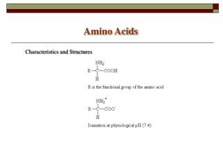
Zwitterion: dipolar form of AA, found at biological pH’s (internal acid/base Rxn)
Formation of Polypeptides
Formation Reaction: • Dehydration reaction • CA + Amine Amide • Amide structure/Peptide bond/Peptide linkage
Amide/Peptide Bonds
Polypeptides: • Small chains of AA (40-50 units) • Many ways to connect together (N!) • ~30 biologically relevant ones • Hormones or Nerve transmitters • Small changes structure HUGE changes in functionality
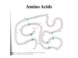
Protein Structure
Proteins – General: • > 50 AA • Linus Pauling – 1954 Nobel Prize α-helix and β-pleated sheet • Fredrick Sanger – 1958 Primary structure of beef insulin
Primary Structure: • #, kind, type, and sequence of AA • Fredrick Sanger (1958 Nobel Prize) Beef Insulin • Several years of work to sequence 51 AA • Hydrolyzed proteins into smaller fragments to analyze • Edman Degradation – split AA at N-Terminal End Gly-Glu-Arg-Gly-Phe-Phe- Fragment 1: Gly-Phe-Phe-Tyr-Thr-Pro-Lys Fragment 2: Gly-Glu-Arg-Gly-Phe-Phe-Tyr-Thr-Pro-Lys Combined: Overlap
Secondary Structure: • Determined by H-bonds between AA-backbone • α-helix AA 4 residues apart, R-groups towards outside • β-pleated sheet AA far apart, R-groups face outwards
Tertiary Structure: • Determined by interactions between R-groups • H-bonds: -COOH and –OH • Ionic/Salt Bridges • Disulfide Bonds • Hydrophobic (form core of protein) • Hydrophilic (face outwards to interact with water)
Quaternary Structure: • Multiple protein units • Non-protein parts • Metal ions • Ex: Hemoglobin • 4 subunits • Fe atoms
Protein Structure Summary
α-helix Structure: • Secondary • Determined by H-bonds between AA-backbone • α-helix AA 4 residues apart, R-groups towards outside
β-pleated sheet structure: • Secondary • Determined by H-bonds between AA-backbone • β-pleated sheet AA far apart, R-groups face outwards
Secondary H-bonds: • Between the C=O and NH of backbone • Responsible for secondary structure • Tertiary H-bonds: • Between the C=O and -NH or -OH of R-groups • Responsible for tertiary structure
Ionic Bonds/Salt Bridges: • Tertiary Structure • Between –COO- and –NH3+ groups
Disulfide bonds: • Tertiary Structure • Between -SH and –SH groups • Mainly between Cys-Cys
Hydrophobic Interactions
Hydrophobic Interactions: • Tertiary Structure • Between –R groups (Alkane and Aromatic) • Interior of proteins to avoid water
Hydrophilic Interactions
Hydrophilic Interactions: • Tertiary Structure • Exterior of proteins to interact with water • Polar groups (OH) • Acidic groups (COOH) • Basic groups (NH2)
Identify 2°/3° Structure
Protein Functions: • Structural Support – skin, connective tissue • Storage – Fe in Liver • Transport – O2 in Hemoglobin • Defense – antibodies, venom • Motion/Movement – muscles • Regulation – blood/glucose/insulin • Catalysis – Enzymes (Ch. 30!)
Denaturation: Loss of 3D conformation in a protein • Disruption of 2°/3°/4° interactions • Does NOT break 1° structure (hydrolysis) • Loss of biological activity • Causes of Denaturation
Xanthoproteic Test
Xanthoproteic Test: • Detects Benzene rings • Yellow color • Phe, Try, Tyr