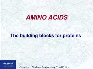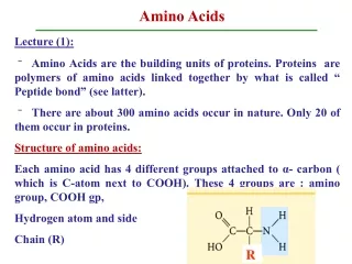
Amino acids
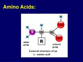
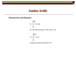
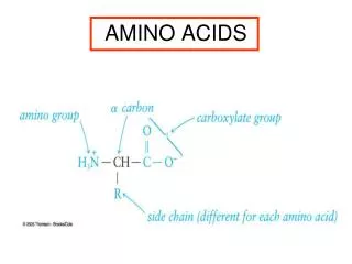
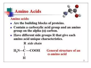
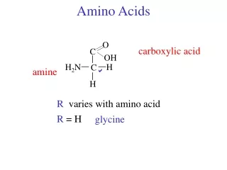
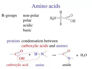
Amino acids. R-groups. non-polar polar acidic basic. proteins . condensation between carboxylic acids and amines. +. +. H 2 O. carboxylic acid. amide. amine. Amides. resonance structure. amides. dipeptide. glycine. alanine. Ala-Gly. +H 2 O. H. H. H. _. _. _. =. =.

Amino acids
E N D
Presentation Transcript
Amino acids R-groups non-polar polar acidic basic proteins condensation between carboxylic acids and amines + + H2O carboxylic acid amide amine
Amides resonance structure amides dipeptide glycine alanine Ala-Gly +H2O
H H H _ _ _ = = = O O O R R R _ _ _ Polypeptides “backbone” _ H N1- C1- C1- N2- C2- C2- N3- C3- C3- OH peptide bonds C-terminal residue N-terminal residue biological activity = structure 4 levels protein structure
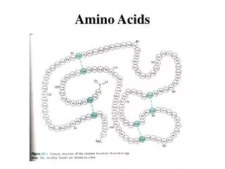
Primary structure sequence of amino acids hemoglobin transports O2 and CO2 300 amino acids 4 protein chains Sickle cell anemia 6thamino acid from N-terminus Glu R Val -CH2CH2-CO2H -CH(CH3)2 water soluble water insoluble
Primary structure study evolution -chain 146 residues horses - humans = 26 pigs - humans = 10 gorillas - humans = 1 1 successful change / 10,000,000 years Primary structure - selective hydrolysis
Phe-Val-Asn-Gln-His Gln-His-Leu-Cys His-Leu-Cys-Gly-Ser His-Leu-Val-Glu Gly-Ser-His-Leu-Val Leu-Val-Glu-Ala Phe-Val-Asn-Gln-His Gln-His-Leu-Cys Leu-Val-Glu-Ala His-Leu-Cys-Gly-Ser Gly-Ser-His-Leu-Val His-Leu-Val-Glu
H H H _ _ _ _ H N1- C1- C1- N2- C2- C2- N3- C3- C3- OH = = = O O O R R R _ _ _ Secondary structure hydrogen bonding backbone groups H-bond donors H-bond acceptors -helix Two main secondary structures: -sheet
H C N = O C = O Alpha helix Every C=O bonded to N-H 4 residues away forms a helix core is backbone R-groups outside 3.6 amino acids per turn proline no H-bonding breaks helix
Beta sheet Every C=O bonded to N-H far apart in 1o structure on different chains peptide chains extended side-by-side maximal H-bonding for anti-parallel chains small R-groups above and below the sheet if not -helix or -sheet random coil
Secondary structure 1o structure some proteins amino acid sequence -helix -sheet 2o structure -sheet silk -helix collagen bone, teeth triple helices keratin hair, skin, wool, hooves cross-linked with disulfide bonds
H H H H C C N N N N C C C C C C S S Disulfide bonds cysteine -CH2-SH S-H H-S reduced [O] oxidized
Protein function enzymes biological catalysts immunoglobulins antibodies transport hemoglobin hormones regulation structural keratin, collagen motion actin, myocin function depends on structure