Pertussis
Pertussis. Highly contagious respiratory infection Classic pertussis, the whooping cough syndrome , usually is caused by B. Pertussis a gram-negative pleomorphic bacillus with fastidious growth requirements

Pertussis
E N D
Presentation Transcript
Highly contagious respiratory infection • Classic pertussis, the whooping cough syndrome, usually is caused by B. Pertussis • a gram-negative pleomorphic bacillus with fastidious growth requirements • Bordetella parapertussis, which causes a similar but milder illness that is not affected by B. pertussis vaccination • Transmission:person to person by coughing. • Adenoviruses have been associated with the pertussis syndrome.
EPIDEMIOLOGY • Outbreaks first described in 16th century • Bordetella pertussis isolated in 1906 • Estimated >500,000 deaths annually worldwide
Pertussis Epidemiology • Reservoir Human Adolescents and adults • Transmission Respiratory droplets Airborne rare
CLINICAL MANIFESTATIONS • Incubation period 3-12 days (up to 21 days) • Insidious onset, similar to minor upper respiratory infection with nonspecific cough • Fever usually minimal throughout course • Apnea & Cyanosis in infant
Pertussis Clinical Features • Catarrhal stage 1-2 weeks • Paroxysmalcough stage 2-4 weeks • Convalescence Weeks to months
Pertussis Clinical Features Stage 1 : Rinorrhea’Lacrimation’Conjectival Injection’Mild cough’’Low grade fever’ Stage 2: Severe cough’Whoop’Cyanosis’Red face’ Postussive Vomiting Stage 3: Decrease Cough & Vomiting’Chronic cough
Young infants may not display the classic pertussis syndrome • the first signs may be episodes of apnea. • Young infants are unlikely to have the classic whoop • are more likely to have CNS damage as a result of hypoxia • Adolescents and adults with pertussis usually present with a prolonged bronchitic
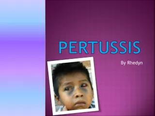
Bilateral scleral hemorrhages and periorbital edema in a young boy with pertussis.
Ecchymoses and conjunctival hemorrhage in a 6-year-old unimmunized child with pertussis
Diagnosis: History &P/E Leuckocytosis (Lymphocytosis) Culture(Nasopharengeal swab) Direct Flurocent Antibody , PCR (Nasopharengeal) Perihilar Infiltration’…signs of segmental lung atelectasis
who has pure or predominant complaint of cough, especially if the following are absent: fever, malaise or myalgia, exanthem or enanthem, sore throat, hoarseness, tachypnea, wheezes, and rales • For sporadic cases, a clinical case definition of cough of ≥14 days' duration with at least 1 associated symptom of paroxysms, whoop, or post-tussive vomiting has a sensitivity of 81% and specificity of 58% for culture confirmation.
Pertussis should be suspected in older children whose cough illness is escalating at 7–10 days and whose coughing episodes are not continuous. • Pertussis should be suspected in infants <3 mo of age with apnea, cyanosis, or an acute life-threatening event (ALTE). B. pertussis is an occasional cause of sudden infant death.
Differential Diagnosis: • Pneumonia • Asthma • RSV, parainfluenza virus, and C. pneumoniae • TB • CF • Foreign body • Bronchiolitis • Mediastinal Lymphadenopathy
COMPLICATIONS • Major complications are most common among infants and young children: hypoxia, apnea, pneumonia, seizures, encephalopathy, and malnutrition • The most frequent complication is pneumonia • Atelectasis may develop secondary to mucous plugs. • The force of the paroxysm may rupture alveoli and produce pneumomediastinum pneumothorax, or interstitial or subcutaneous emphysema • epistaxis; hernias; and retinal and subconjunctival hemorrhages
Treatment • Erythromycin 50 mg/kg/D PO (14 DAY) • Azithromycin , clarithromycin ,TMP-SMZ • Salbutamol • Moist O2 • Nasopharengeal suction • IV Fluid • No Immunoglobulin’No Antitussive drugs’No Steroid
PREVENTION • Pertussis vaccine has an efficacy of 70% to 90% • Erythromycin and other macrolides are effective in preventing disease in contacts exposed to pertussis • Close contacts younger than 7 years should receive a booster dose of DTaP also should be given a macrolide antibiotic. • Close contacts older than age 7 should receive prophylactic macrolide antibiotic for 10 to 14 days, but not the vaccine