Understanding Sensation: How Our Brain Interprets Environmental Stimuli
Sensation is the process by which the brain receives information about the environment and the body through specialized receptors. These receptors transduce various forms of energy into nerve impulses that are then conducted to the cerebral cortex for interpretation. Sensory modalities include touch, pain, taste, smell, sight, hearing, and balance. Different types of receptors, such as mechanoreceptors and nociceptors, allow us to perceive different stimuli. This guide explores the classifications of sensory receptors, the nature of sensations, and how our brain interprets them.

Understanding Sensation: How Our Brain Interprets Environmental Stimuli
E N D
Presentation Transcript
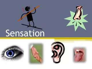
Sensation • Senses: Means by which brain receives information about environment and body • General: Distributed over large part of body • Somatic: Touch, pressure, temperature, proprioception, pain • Visceral: Internal organs and consist mostly of pain and pressure • Special senses: Smell, taste, sight, hearing, balance • Sensation or perception: Conscious awareness of stimuli received by sensory receptors
Sensation • Sensation or perception is the conscious awareness of stimuli received by receptor. • Receptors transduce (change) different forms of energy into nerve impulses • Nerve impulses are conducted to the brain • Stimulus must initiate and action potential in the cerebral cortex • The brain interprets these impulses as sound or sight even though the impulses themselves are identical in nature. • The cerebral cortex screens the information and ignores most of what it receives – subconscious • Our senses act as energy filters that perceive a narrow range of energy.
Sensation Requires: • A stimulus • Activation of a receptor, • Conduction of an action potential to a specific region of the CNS • Translation or interpretation of the signal. • Sensation or awareness of a stimuli occurs in the cerebral cortex.
General Properties of Receptors • A receptor is any structure specialized to detect a stimulus. • All receptors are transducers, changing stimulus energy into nerve energy. • Sensory receptors transmit four kinds of information: • Modality refers to the type of stimulus or sensation it produces (vision, hearing, taste, etc.). • Location is also indicated by which nerve fibers are firing. • Sensory projection is the ability of the brain to identify the site of stimulation. • Intensity of stimulous • Duration is encoded in the way nerve fibers change their firing frequencies over time. • Tonic vs Phasic receptor adaptation.
Types of Sensory Receptors • Classification by Stimulus Modality • Mechanoreceptors: Compression, bending, stretching of cells • Chemoreceptors: Smell and taste • Thermoreceptors: Temperature • Photoreceptors: Light as vision • Nociceptors: Pain • Classification by Origin of Stimuli • Exteroreceptors: Associated with skin • Visceroreceptors: Associated with organs • Proprioceptors: Associated with joints, tendons
Sensory Nerve Endings • Unencapsulated Nerve Endings • Free nerve endings: Cold receptors and warm • Merkel’s disk: Light touch, superficial pressure • Hair follicle receptor: Light touch, bending of hair
Sensory Nerve Endings in SkinEncapsulated Nerve Endings • Pacinian corpuscle: Deep cutaneous pressure, vibration and proprioception • Meissner’s corpuscle: Two-point discrimination • Ruffini’s end organ: Continuous touch or pressure • Muscle spindle: Proprioception as to muscle stretch and control of muscle tone • Golgi tendon organ: Important in muscle contraction and tendon stretch proprioception
Responses of Sensory Receptors • Receptor: Interaction of stimulus with sensory receptor produces a local potential • Primary: Have axons that conduct action potential in response to receptor potential • Secondary: Have no axons and receptor potentials produced do not result in action potentials but cause release of neurotransmitters • Accommodation or adaptation: Decreased sensitivity to a continued stimulus • Proprioceptors • Tonic: Example is know where little finger is without looking • Phasic: Example is you know where hand is as it moves
Sensory Nerve Tracts • Transmit action potentials from periphery to brain • Each pathway involved with specific modalities • First half of word indicates origin, second half indicates termination
Spinothalamic System • Conveys cutaneous sensory information to brain • Unable to localize source of stimulus • Divisions • Lateral for pain and temperature • Anterior for light touch, pressure, tickle, itch
Dorsal-Column/Medial-Lemniscal System • Carries sensations of • Two-point discrimination • Proprioception • Pressure • Vibration • Tracts • Fasciculus gracilis • Fasciculus cuneatus
Spinocerebellar System • Carry proprioceptive information to cerebellum • Actual movements can be monitored and compared to cerebral information representing intended movement • Tracts • Posterior • Anterior
Pain • Types • Referred: Sensation in one region of body that is not source of stimulus • Phantom: Occurs in people who have appendage amputated or structure removed as tooth • Chronic: Not a response to immediate direct tissue injury
Special Senses • Olfaction • Taste • Visual system • Hearing and balance
Olfaction • Sense of smell • Olfactory epithelium • 10-20 million neurons • Bipolar neurons project through cribiform plate. • Olfactory hairs • 10 – 20 Cilia per neuron. • Embedded in a mucous layer • Only neurons exposed to external environment • Replaced every 60 days.
Olfactory Physiology • Process of Olfaction • Airborne chemicals are dissolved in the fluid covering the olfactory epithelium. • Chemicals must be volatile and water soluble. • Odor molecule binds with a specific receptor • G-protein coupled membrane receptor) • a second messenger is produced, • Sodium channels are opened in the membrane. • The cell is depolarized creating an axon potential. • Olfactory Discrimination • We can discriminate between ~10,000 different odors. • There are between 7-50 primary classes of odors • A characteristic fingerprint of the odor is used to identify the odor. • Olfactory receptors adapt quickly • Some odors can stimulate nociceptors in the trigeminal nerve.
Olfactory Neuronal Pathways and the Cortex • Olfactory neurons project to the olfactory bulb. • Mitral cells project to the olfactory cortex. • Lateral olfactory area: conscious perception of smell • Medial olfactory area: visceral and emotional reactions • Intermediate olfactory area merges information from medial and lateral areas and projects back to olfactory bulb to modulate neuronal activity there.
Clinical Considerations of Olfaction • Anosmia – inability to smell (1.2% of the population) • Ability to smell decreases with age. • 98-99% of people can smell banana, rose and cloves. • 35% of the population cannot smell androstenone (body odor).
Papillae and Taste Buds • Taste results from the action of chemicals on the taste buds found on papillae. • ~10,000 taste buds • Papillae Types • Circumvallate • Fungiform • Foliate • Filiform • Taste Bud Structure • Supporting cells - Form an exterior supporting capsule • Gustatory or taste cells contain gustatory villi or hairs with surface receptors and are replaced every 7 to 10 days.
Physiology of Taste • Process of Taste • Molecules are dissolved in saliva. • Substance enters taste pore and attaches to chemoreceptor molecule • Depolarization of the taste cell. • Taste cells have no axons but release neurotransmitter • Neurotransmitter stimulates action potential in cells associated with the gustatory cells.
Four Primary Taste Sensations Exist: • Salty • lateral anterior of tongue • The presence of Na+ is detected • Sweet • tip of the tongue • most organic molecules (particularly sugars) are sweet. • Sour • posterior lateral portion of the tongue • H+ are detected • all acids taste sour. • Bitter • most posterior central portion of the tongue • most sensitive • protective function - most poisons are bitter • Umami (Glutamate) may also be considered.
Fibrous tunic: Outer Sclera: White outer layer, maintains shape, protects internal structures, provides muscle attachment point, continuous with cornea Cornea: Avascular, transparent, allows light to enter eye and bends and refracts light Vascular tunic: Middle Iris: Controls light entering pupil; smooth muscle Ciliary muscles: Control lens shape; smooth muscle Retina: Inner Contains neurons sensitive to light Macula lutea or fovea centralis: Area of greatest visual acuity Optic disc: Blind spot Compartments Anterior: Aqueous humor Posterior: Vitreous humor Lens Held by suspensory ligaments attached to ciliary muscles Transparent, biconvex Anatomy of the Eye
Compartments of the Eye • Posterior Compartment • Vitreous Humor • Anterior Compartment • Anterior Chamber • Posterior Chamber • Aqueous Humor • Produced by ciliary processes
Functions of the Complete Eye • Eye functions like a camera • Iris allows light into eye • Lens, cornea, humors focus light onto retina • Light striking retina is converted into action potentials relayed to brain
Light • Focal point: Point where light rays converge and cross • The more spherical the lens the more the light is bent. • Reflection: light rays bounce off a non transparent object • Visible light: Portion of electromagnetic spectrum detected by human eye • The visible spectrum ranges form ~400 to 700 nm • Refraction: Bending of light • Divergence: Light striking a concave surface • Convergence: Light striking a convex surface • The cornea, aqueous humor, lens and vitreous humor all refract light.
Focus and Accommodation • Focusing system of the eye creates a clear image on the retina. • Emmetropia: Normal resting condition of lens • Far vision: 20 feet + from eye. • Near vision: Closer than 20 feet • Accommodation • Occurs via changes in the shape of the lens. • Pupil constriction • Depth of focus • Convergence • The inverted image on the retina is detected by photoreceptors and passed via action potentials to the visual cortex.