Download
1 / 7
70 likes | 139 Vues
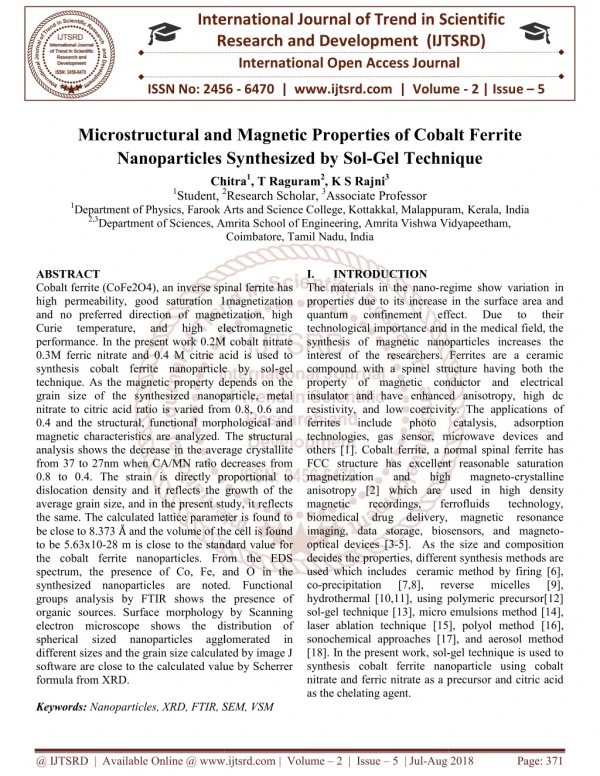
Cobalt ferrite CoFe2O4 , an inverse spinal ferrite has high permeability, good saturation 1magnetization and no preferred direction of magnetization, high Curie temperature, and high electromagnetic performance. In the present work 0.2M cobalt nitrate 0.3M ferric nitrate and 0.4 M citric acid is used to synthesis cobalt ferrite nanoparticle by sol gel technique. As the magnetic property depends on the grain size of the synthesized nanoparticle, metal nitrate to citric acid ratio is varied from 0.8, 0.6 and 0.4 and the structural, functional morphological and magnetic characteristics are analyzed. The structural analysis shows the decrease in the average crystallite from 37 to 27nm when CA MN ratio decreases from 0.8 to 0.4. The strain is directly proportional to dislocation density and it reflects the growth of the average grain size, and in the present study, it reflects the same. The calculated lattice parameter is found to be close to 8.373 u00c3u2026 and the volume of the cell is found to be 5.63x10 28 m is close to the standard value for the cobalt ferrite nanoparticles. From the EDS spectrum, the presence of Co, Fe, and O in the synthesized nanoparticles are noted. Functional groups analysis by FTIR shows the presence of organic sources. Surface morphology by Scanning electron microscope shows the distribution of spherical sized nanoparticles agglomerated in different sizes and the grain size calculated by image J software are close to the calculated value by Scherrer formula from XRD. Chitra | T Raguram | K S Rajni "Microstructural and Magnetic Properties of Cobalt Ferrite Nanoparticles Synthesized by Sol-Gel Technique" Published in International Journal of Trend in Scientific Research and Development (ijtsrd), ISSN: 2456-6470, Volume-2 | Issue-5 , August 2018, URL: https://www.ijtsrd.com/papers/ijtsrd15871.pdf Paper URL: http://www.ijtsrd.com/physics/other/15871/microstructural-and-magnetic-properties-of-cobalt-ferrite-nanoparticles-synthesized-by-sol-gel-technique/chitra<br>

E N D
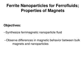
International Research Research and Development (IJTSRD) International Open Access Journal nd Magnetic Properties of Cobalt Ferrite Nanoparticles Synthesized by Sol-Gel Technique Chitra1, T Raguram2, K S Rajni3 2Research Scholar, 3Associate Professor of Physics, Farook Arts and Science College, Kottakkal, Malappuram, of Sciences, Amrita School of Engineering, Amrita Vishwa Vidyapeetham, Coimbatore, Tamil Nadu, India International Journal of Trend in Scientific Scientific (IJTSRD) International Open Access Journal ISSN No: 2456 ISSN No: 2456 - 6470 | www.ijtsrd.com | Volume 6470 | www.ijtsrd.com | Volume - 2 | Issue – 5 Microstructural and Magnetic Properties Nanoparticles Synthesized f Cobalt Ferrite Gel Technique Chitra 1Student, 2 1Department of Physics, Farook Arts and Science College, Kott 2,3Department of Sciences, Amrita S akkal, Malappuram,Kerala,India Amrita Vishwa Vidyapeetham, ABSTRACT Cobalt ferrite (CoFe2O4), an inverse spinal ferrite has high permeability, good saturation 1magnetization and no preferred direction of magnetization, high Curie temperature, and performance. In the present work 0.2M cobalt nitrate 0.3M ferric nitrate and 0.4 M citric acid is used to synthesis cobalt ferrite nanoparticle by sol technique. As the magnetic property depends on the grain size of the synthesized nanoparticle, metal nitrate to citric acid ratio is varied from 0.8, 0.6 and 0.4 and the structural, functional morphological and magnetic characteristics are analyzed. The structural analysis shows the decrease in the average crystallite from 37 to 27nm when CA/MN ratio decreases from 0.8 to 0.4. The strain is directly proportion dislocation density and it reflects the growth of the average grain size, and in the present study, it reflects the same. The calculated lattice parameter is found to be close to 8.373 Å and the volume of the cell is found to be 5.63x10⁻²⁸ m is close to the standard value for the cobalt ferrite nanoparticles. From the EDS spectrum, the presence of Co, Fe, and O in the synthesized nanoparticles are noted. Functional groups analysis by FTIR shows the presence of organic sources. Surface morphology by Sc electron microscope shows the distribution of spherical sized nanoparticles different sizes and the grain size calculated by image J software are close to the calculated value by Scherrer formula from XRD. Keywords: Nanoparticles, XRD, FTIR, SEM, VSM I. The materials in the nano-regime show variation in properties due to its increase in the surface area and quantum confinement technological importance and in the medical field, the synthesis of magnetic nanoparticles increases the interest of the researchers. Ferrites are a ceramic compound with a spinel structure having both the property of magnetic conductor and electrical insulator and have enhanced anisotropy, high dc resistivity, and low coercivity. The applications of ferrites include photo technologies, gas sensor, microwave devices and others [1]. Cobalt ferrite, a normal spinal ferrite has FCC structure has excellent reasonable saturation magnetization and high anisotropy [2] which are u magnetic recordings, biomedical drug delivery, magnetic resonance imaging, data storage, biosensors, and magneto optical devices [3-5]. As the size and composition decides the properties, different synthesis met used which includes ceramic method by firing [6], co-precipitation [7,8], hydrothermal [10,11], using polymeric precursor[12] sol-gel technique [13], micro emulsions laser ablation technique [15], polyol method sonochemical approaches [17], and aerosol method [18]. In the present work, sol- synthesis cobalt ferrite nanoparticle using cobalt nitrate and ferric nitrate as a precursor and citric acid as the chelating agent. INTRODUCTION Cobalt ferrite (CoFe2O4), an inverse spinal ferrite has high permeability, good saturation 1magnetization and no preferred direction of magnetization, high Curie temperature, and performance. In the present work 0.2M cobalt nitrate M ferric nitrate and 0.4 M citric acid is used to synthesis cobalt ferrite nanoparticle by sol-gel technique. As the magnetic property depends on the grain size of the synthesized nanoparticle, metal nitrate to citric acid ratio is varied from 0.8, 0.6 and 0.4 and the structural, functional morphological and magnetic characteristics are analyzed. The structural analysis shows the decrease in the average crystallite from 37 to 27nm when CA/MN ratio decreases from 0.8 to 0.4. The strain is directly proportional to dislocation density and it reflects the growth of the average grain size, and in the present study, it reflects the same. The calculated lattice parameter is found to and the volume of the cell is found regime show variation in properties due to its increase in the surface area and quantum confinement technological importance and in the medical field, the synthesis of magnetic nanoparticles increases the rest of the researchers. Ferrites are a ceramic compound with a spinel structure having both the property of magnetic conductor and electrical insulator and have enhanced anisotropy, high dc resistivity, and low coercivity. The applications of photo technologies, gas sensor, microwave devices and others [1]. Cobalt ferrite, a normal spinal ferrite has FCC structure has excellent reasonable saturation magnetization and high anisotropy [2] which are used in high density magnetic recordings, biomedical drug delivery, magnetic resonance imaging, data storage, biosensors, and magneto- 5]. As the size and composition decides the properties, different synthesis methods are used which includes ceramic method by firing [6], precipitation [7,8], hydrothermal [10,11], using polymeric precursor[12] micro emulsions method [14], laser ablation technique [15], polyol method [16], sonochemical approaches [17], and aerosol method effect. effect. Due Due to to their their high high electromagnetic electromagnetic catalysis catalysis, adsorption magneto magneto-crystalline ferrofluids ferrofluids technology, technology, to the standard value for the cobalt ferrite nanoparticles. From the EDS spectrum, the presence of Co, Fe, and O in the synthesized nanoparticles are noted. Functional groups analysis by FTIR shows the presence of organic sources. Surface morphology by Scanning electron microscope shows the distribution of spherical sized nanoparticles different sizes and the grain size calculated by image J software are close to the calculated value by Scherrer reverse reverse micelles micelles [9], [9], agglomerated agglomerated in in -gel technique is used to synthesis cobalt ferrite nanoparticle using cobalt nitrate and ferric nitrate as a precursor and citric acid RD, FTIR, SEM, VSM @ IJTSRD | Available Online @ www.ijtsrd.com @ IJTSRD | Available Online @ www.ijtsrd.com | Volume – 2 | Issue – 5 | Jul-Aug 2018 Page: 371
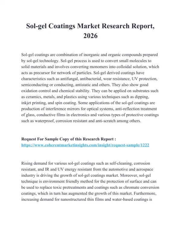
International Journal of Trend in Scientific Research and Development (IJTSRD) ISSN: 2456-6470 Where M is the molecular weight of the sample and A is the Avogadro’s number (6.022x10²³mol-1) and ‘a’ is the lattice parameter The X-ray diffraction data is used to calculate ionic radii (rA, rB) and bond lengths (A-O), (B-O) at the tetrahedral and octahedral sites, is given by the equations. rA = (u-¼)a√3- r(O⁻²) Å rB = (⁵?₈-u)a- r(O⁻²) Å A-O = (u-¼)a√3 Å B-O= (⁵?₈-u)a Å Where ‘a’ is the lattice constant; r(O⁻²) is the radius of oxygen ion (1.35 Å); u is the oxygen ion parameter and for ideal spinel ferrite u=3/8. Hopping lengths at tetrahedral sites (Lᴀ) and at octahedral sites (Lв) which is the distance between the magnetic ions is calculated by the following equation Lᴀ=a(√³?₄) Å Lв=a(√²?₄) Å III. RESULTSANDDISCUSSION All paragraphs must be indented. All paragraphs must be justified, i.e. both left-justified and right- justified. A.Structural Analysis II. A.Cobalt Ferrite Nanoparticles Synthesis Cobalt ferrite nanoparticles are synthesized using citric acid as a precursor. 0.2M Cobalt nitrate [Co(NO3)2•6H2O] and [Fe(NO3)3•9H2O] and 0.4 M citric acid [C7H8O7.H2O] is prepared in distilled water and the concentration of citric acid (CA) to metal nitrate (MN) solution is taken as 0.8, 0.6 and 0.4. The pH of the solution is maintained at 8. The mixed solution is then heated to 80 °C with constant stirring for two hours till a brown gel is obtained. The gel is heated to 800 °C for three hours to remove excess of water. During the process of drying, the gel swells into a fluffy mass and eventually broke into brittle flakes. B.Characterisation The samples were subjected to Powder X-ray diffraction analysis using Shimadzu XRD 6000 diffractometer with CuKα radiation of wavelength 1.541 Å. The functional group is analyzed by FTIR using Perkin-Elmer spectrometer by KBr pellet technique in the range of 4000-400 cm-1. The morphology analysis is assessed by Scanning Electron microscopy using JEOL (JSM 6390). Magnetic properties of the samples are analyzed by VSM (Model 7407) at room temperature with the maximum applied field of 15kOe. The crystallite size (D) is calculated using the Scherrer formula [19] from the full- width half maximum (FWHM) (β) for the most intense peak (311) D= (k λ) / (β cosθ) Å Miller indices (h, k, l) are related to inter-atomic spacing (d) spacing and for cubic crystals, the lattice parameter ‘a’ is calculated for prominent peak (311) using the relation [20] a = dhkl /(h2 + k2 + l2)1/2Å The strain (ε) is calculated from the relation with β ε = β cosθ / 4 The dislocation density (ρ) is defined as the length of dislocation lines per unit volume of the crystal, is calculated from the formula ρ=1/D2 lines /cm3 The theoretical X-ray density, (ρx) is calculated by the relation [20] ρx =8M/Na3 g/cm3 EXPERIMENTAL 0.2M ferric nitrate (6) (7) (8) (9) (10) (11) (1) (2) (3) Fig. 1 PXRD Pattern of cobalt ferrite nanoparticle with different CA/MN ratio The Structural studies are carried out and PXRD pattern as shown in the fig.1. The observed peaks (220), (311), (400), (440) and (511) matches well with the JCPDS data of cobalt ferrite nanoparticle [21]. It is noted that the slight decrease in the intensity of the plane corresponding to (311) compared to (400) plane (4) (5) @ IJTSRD | Available Online @ www.ijtsrd.com | Volume – 2 | Issue – 5 | Jul-Aug 2018 Page: 372
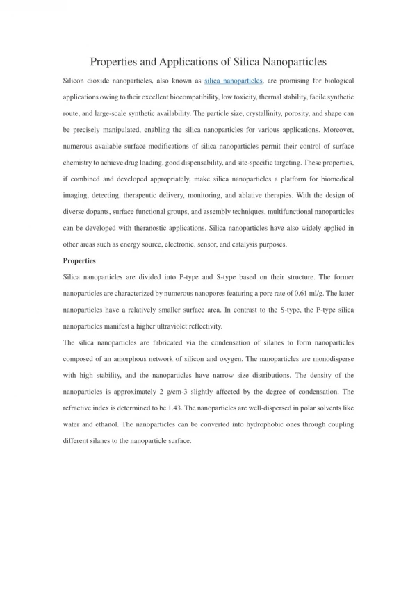
International Journal of Trend in Scientific Research and Development (IJTSRD) ISSN: 2456 International Journal of Trend in Scientific Research and Development (IJTSRD) ISSN: 2456 International Journal of Trend in Scientific Research and Development (IJTSRD) ISSN: 2456-6470 is observed when the CA/MN ratio decreases from 0.8 to0.4. The microstructural parameters of cobalt ferrite nanoparticle are shown in table 1. It is noted that the grain size varies from 37 to 27nm as the ratio of CA/MN decreases from 0.8 to 0.4 is attributed to the burning of nitrates due the higher volum concentration of the chelating agent. The variation in the strain and the dislocation density with respect to crystallite size. Fig. 2 shows the lattice constant and X-ray density varies with crystallite size. The observed lattice constant is found to be less than the value for the bulk (8.373 Å) is attributed to the nanosizing effect. It is noted that the lattice constant decreases from 8.314 to 8.170 when CA /MN decreases from0.8 to 0.4. The calculated volume of the cell is less compared to the bulk value (590.99A°) is attributed to the nanosizing effect in cobalt ferrite is observed when the CA/MN ratio decreases from 0.8 to0.4. The microstructural parameters of cobalt ferrite nanoparticle are shown in table 1. It is noted that the grain size varies from 37 to 27nm as the ratio of CA/MN decreases from 0.8 to 0.4 is attributed to the burning of nitrates due the higher volume and concentration of the chelating agent. The variation in the strain and the dislocation density with respect to crystallite size. Fig. 2 shows the lattice constant and ray density varies with crystallite size. The nanoparticles [22]. X-ray density found to increase with a decrease in the lattice constant and similar type of observations are noted for Mg prepared at different molar concentrations [23]. Also, the value of the X-ray density is higher than their bulk value is due to the formation of pores during the synthesis process and due to the ionic radii [13, 24]. The mean ionic radius of octahedral site B (rB) is found to decrease slowly than the tetrahedral site A (rA). A similar type of observations is noted by other investigators for their Co- hopping length LA and LB, bond length A decrease gradually with the decrease in the CA/MN ratio and the crystallite size which reflects the decrease in lattice constant which is shown in the table 1. ray density found to increase with a decrease in the lattice constant and similar type of observations are noted for Mg-Zn ferrite system molar concentrations [23]. Also, ray density is higher than their bulk value is due to the formation of pores during the synthesis process and due to the ionic radii [13, 24]. The mean ionic radius of octahedral site B (rB) is decrease slowly than the tetrahedral site A (rA). A similar type of observations is noted by other o be less than the -Zn system [25]. The value for the bulk (8.373 Å) is attributed to the nanosizing effect. It is noted that the lattice constant decreases from 8.314 to 8.170 when CA /MN decreases from0.8 to 0.4. The calculated volume of hopping length LA and LB, bond length A-O and B-O decrease gradually with the decrease in the CA/MN the crystallite size which reflects the decrease in lattice constant which is shown in the value (590.99A°) is attributed to the nanosizing effect in cobalt ferrite 38 Crystallite Size 36 Crystallite Size (nm) 34 32 30 28 26 0.4 0.5 0.6 (a) 0.7 0.8 CA/MN (b) Fig. 2 Structural analysis of cobalt ferrite nanoparticle a) Crystallite size Vs CA/MN ratio a) Crystallite size Vs CA/MN ratio b) Lattice constants, X-Ray Density Vs Crystallite size ratio Fig. 2 Structural analysis of cobalt ferrite nanoparticles with different CA/MN ratio s with different CA/MN ratio Ray Density Vs Crystallite size ratio Table I Hopping Length, Bond Length and Radius of The Ions a I Hopping Length, Bond Length and Radius of The Ions at The Tetrahedral and Octahedral Sites o Cobalt Ferrite Nanoparticles trahedral and Octahedral Sites of (rA) (x10-11m) 4.3927 4.3587 4.3277 (rB) (x10-11m) 7.1607 7.1214 7.0856 CA/MN 0.8 0.6 0.4 LA(A°) 3.0991 3.0932 3.0878 LB(A°) 2.9218 2.9163 2.9112 2.9112 (A°) A-O (A°) 1.7892 1.7858 1.7827 B-O (A°) 2.0660 2.0621 2.0585 2.9218 2.9163 B.Functional Group Analysis Fig.3 Structural analysis of cobalt ferrite nanoparticles with different CA/MN ratio CA/MN ratio. b) Lattice constants, X Crystallite size ratio Fig. 3 shows the FTIR Spectra of the prepared samples. The peak positions of are listed in table 2. It is noted that Nitrate group has six normal vibrations that are IR active and found in IR spectra. They are (i) anti IR spectra. They are (i) anti-symmetric stretching Structural analysis of cobalt ferrite nanoparticles with different CA/MN ratio. a) Crystallite size Vs Lattice constants, X-Ray Density Vs Crystallite size ratio Fig. 3 shows the FTIR Spectra of the prepared samples. The peak positions of the functional groups are listed in table 2. It is noted that Nitrate group has six normal vibrations that are IR active and found in @ IJTSRD | Available Online @ www.ijtsrd.com @ IJTSRD | Available Online @ www.ijtsrd.com | Volume – 2 | Issue – 5 | Jul-Aug 2018 Page: 373
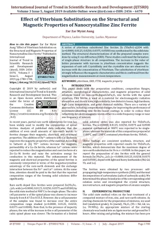
International Journal of Trend in Scientific Research and Development (IJTSRD) ISSN: 2456 International Journal of Trend in Scientific Research and Development (IJTSRD) ISSN: 2456 International Journal of Trend in Scientific Research and Development (IJTSRD) ISSN: 2456-6470 (1629 cm-1),(ii) symmetric stretching (1388 cm 1),(iii) totally symmetric stretching (1052 cm out of plane bending (870 cm-1),(v) anti in-plane bending (729 cm-1),(vi) symmetric in bending (664 cm-1). From the spectra, it is noted that the peak observed at 600 cm-1 corresponds to characteristic stretching of Fe-O [26]. The peaks 1),(ii) symmetric stretching (1388 cm- ic stretching (1052 cm-1),(iv) 1),(v) anti-symmetric 1),(vi) symmetric in-plane 1). From the spectra, it is noted that corresponds to organic sources are also exists in the compound which is observed in the XRD. The peak positions of the functional groups are listed in the table and the presence of organic sources that exists in n be removed by annealing the samples at suitable temperature and duration. corresponds to organic sources are also exists in the compound which is observed in the XRD. The peak positions of the functional groups are listed in the table and the presence of organic sources that exists in the compound can be removed by annealing the samples at suitable temperature and duration. 1 corresponds to O [26]. The peaks TABLE II FT-IR Peak positions of cobalt ferrite nanoparticles Wavenumbers (cm-1) 0.8 0.6 0.4 3428.92 3436.43 3435.64 2923.44 2923.31 2923.14 IR Peak positions of cobalt ferrite nanoparticles Spectral Assignments stretching vibrations of free and absorbed water[28] axial deformation of C-H bond (antisymmetric and symmetric stretching)[31] axial deformation of C-H bond(antisymmetric and symmetric stretching)[31] C=C vibrations H-OH bending of water molecule [28,29] symmetric stretching Nitrate anion overlap with vibrations of C totally symmetric stretching Intrinsic stretching vibration of bond between oxygen and tetrahedral stretching vibrations of free and absorbed water[28] H bond (antisymmetric and symmetric stretching)[31] 2849 2849 2849 stretching)[31] 1730 1625.98 1390.82 1242 1081 595.62 1730 1626.33 1382.28 1242 1081 587.67 - 1611.80 1383.46 - 1081 584.96 OH bending of water molecule [28,29] Nitrate anion overlap with vibrations of C-H [30] stretching Intrinsic stretching vibration of bond between oxygen and tetrahedral Intrinsic stretching vibration of bond between oxygen and tetrahedral metal ion Mtetra↔O[32] Octahedral metal stretching[32] Octahedral metal stretching[32] 461.105 457.884 438.221 C. Morphological Analysis The scanning electron microscopic images of SEM images show a uniform distribution of grain with agglomeration in the nanometric region confirming the crystalline nature of the particle. It is also noted that at higher CA/MN ratio, gas evolution during the synthesis process at a higher temperature. The grain size calculated by image J software is found to be close with the grain size calculated from Scherrer formula form XRD. (a)CA/MN=0.8 (b) CA/MN =0.6 (c) CA/MN =0.4 (a)CA/MN=0.8 (b) CA/MN =0.6 (c) CA/MN =0.4 Fig. 4 Morphological analysis of cobalt ferrite nanoparticles with different CA/MN ratio Fig. 4 Morphological analysis of cobalt ferrite nanoparticles with different CA/MN ratio The scanning electron microscopic images of the synthesized samples are shown in Fig. 4. It is evident that the SEM images show a uniform distribution of grain with agglomeration in the nanometric region confirming the crystalline nature of the particle. It is also noted that at higher CA/MN ratio, pores are noted which is due to the gas evolution during the synthesis process at a higher temperature. The grain size calculated by image J software is found to be close with the grain size calculated from Scherrer formula form XRD. software is found to be close with the grain size calculated from Scherrer formula form XRD. the synthesized samples are shown in Fig. 4. It is evident that the SEM images show a uniform distribution of grain with agglomeration in the nanometric region confirming the pores are noted which is due to the gas evolution during the synthesis process at a higher temperature. The grain size calculated by image J (a)CA/MN=0.8 (b) CA/MN =0.6 (c) CA/MN =0.4 Fig. 4 Morphological analysis of cobalt ferrite nanoparticles with different CA/MN ratio @ IJTSRD | Available Online @ www.ijtsrd.com @ IJTSRD | Available Online @ www.ijtsrd.com | Volume – 2 | Issue – 5 | Jul-Aug 2018 Page: 374
International Journal of Trend in Scientific Research and Development (IJTSRD) ISSN: 2456 International Journal of Trend in Scientific Research and Development (IJTSRD) ISSN: 2456 International Journal of Trend in Scientific Research and Development (IJTSRD) ISSN: 2456-6470 D.Compositional Analysis Fig. 5 shows the compositional analysis by Energy dispersive X the synthesized sample is CoFe2O4 and the atomic ration of Co:Fe:O is close to that of CoFe atomic weight (%) of the prepared samples are shown in the fig. 6. (a) Fig. 5 Compositional analysis of cobalt ferrite nanoparticle with different CA/MN ratio O (k) Fe (K) Co (K) ositional analysis by Energy dispersive X-ray spectroscopic (EDS). The analyses indicate and the atomic ration of Co:Fe:O is close to that of CoFe atomic weight (%) of the prepared samples are shown in the fig. 6. ray spectroscopic (EDS). The analyses indicate and the atomic ration of Co:Fe:O is close to that of CoFe2O4 formula. The (b) (c) Fig. 5 Compositional analysis of cobalt ferrite nanoparticle with different CA/MN ratio Fig. 5 Compositional analysis of cobalt ferrite nanoparticle with different CA/MN ratio than the A site. This compressive strain leads to the breaking of surface exchange bonds which results in canted spin structure. This kind of structure weakens AA, BB and AB interactions which results in low magnetization values at room temperature compared to the bulk cobalt ferrite particle [32]. Fig. 7 (a) and (b) shows the VSM analysis and Coercivity and Saturation Magnetization varies with the crystallite size. It is noted that the coercivity and saturation magnetization increases with the decrease in the average crystallite size and CA/MN ratio. The coercivity increase from 36 to 112 when the crystallite size decreases from 38 to 27 nm. The decrease in coercivity is due to the expected crossover from a single domain to multidomain as particle size increases from 27 to 38nm [33]. The existence of the superparamagnetic behaviour of the synthesised particle is attributed to the finite crystallite size and the shape of the material [34]. The saturation magnetisation is found to be lesser than the saturation magnetisation for bulk (80 emu/g) is attributed to the surface defects and morphology [35]. The surface defects are due to the finite crystallite size which leads to the non-collinear magnetic moments on the surface of the nanoparticle. These type of defects are intense in the ferromagnetic system and the superexchange interaction occurs through the oxygen ion [36]. The experimental magnetic moment (ηB) is calculated by the relation [37] ηB = MMS/ 5585 Where M is the molecular weight. Where M is the molecular weight. compressive strain leads to the breaking of surface exchange bonds which results in canted spin structure. This kind of structure weakens AA, BB and AB interactions which results in low magnetization values at room temperature compared errite particle [32]. Fig. 7 (a) and (b) shows the VSM analysis and Coercivity and Saturation Magnetization varies with the crystallite size. It is noted that the coercivity and saturation magnetization increases with the decrease in the e size and CA/MN ratio. The coercivity increase from 36 to 112 when the crystallite size decreases from 38 to 27 nm. The decrease in coercivity is due to the expected crossover from a single domain to multidomain as particle size 40 35 30 Atomic Weight (%) 25 20 15 10 5 0 0.8 0.4 0.6 CA/MN Fig. 6 Atomic Weight (%) of cobalt ferrite nanoparticle with different CA/MN ratio nanoparticle with different CA/MN ratio Fig. 6 Atomic Weight (%) of cobalt ferrite [33]. The existence of the superparamagnetic behaviour of the synthesised particle is attributed to the finite crystallite size and the shape of the material [34]. The saturation magnetisation is found to be lesser than the saturation k (80 emu/g) is attributed to the surface defects and morphology [35]. The surface defects are due to the finite crystallite size which collinear magnetic moments on the surface of the nanoparticle. These type of defects are ferromagnetic system and the n occurs through the oxygen E. VSM Analysis The magnetic parameters are analyzed by vibrating sample magnetometer (VSM) with a maximum field of 15kOe at room temperature. The properties of cobalt ferrite nanoparticles are decided by cation distribution, size and the synthesis procedure [19, 20] and these properties are explained on the basis of the surface disorder. The random distribution of cation and the canted spi results in surface disorder. Also, broken exchange bonds and high anisotropy on the surface leads to surface disorder [27, 28]. In the case of cobalt ferrite nanoparticle Co2+ ion arbitrarily occupy both tetrahedral (A) site and octahedral (B) of the Fe3+ ion (0.645 Å) move to octahedral site to replace heavier Co2+ion (0.72 Å) [29 migration of Fe3+ ion to B site results in compressive strain as the distance between the B site is smaller strain as the distance between the B site is smaller The magnetic parameters are analyzed by vibrating sample magnetometer (VSM) with a maximum field of 15kOe at room temperature. The magnetic properties of cobalt ferrite nanoparticles are decided by cation distribution, size and the synthesis procedure [19, 20] and these properties are explained on the basis of the surface disorder. The random distribution of cation and the canted spin structure results in surface disorder. Also, broken exchange bonds and high anisotropy on the surface leads to surface disorder [27, 28]. In the case of cobalt ferrite ion arbitrarily occupy both tetrahedral (A) site and octahedral (B) sites, so some ) move to octahedral site to The experimental magnetic moment (ηB) is calculated ) [29-31].The migration of Fe3+ ion to B site results in compressive @ IJTSRD | Available Online @ www.ijtsrd.com @ IJTSRD | Available Online @ www.ijtsrd.com | Volume – 2 | Issue – 5 | Jul-Aug 2018 Page: 375
International Journal of Trend in Scientific Research and Development (IJTSRD) ISSN: 2456-6470 The anisotropy constant is evaluated using following relation [38] Hc = 0.96 K/ MS, Where K is anisotropy constant Table 3 shows the variation of magnetic moment, remanence ratio and anisotropic constant of the cobalt ferrite nanoparticle. The variation of magnetic moment with grain size can be explained on the basis of cation distribution and the strength of the super- exchange interaction between the ions on the tetrahedral (A) and octahedral (B) sublattices. The remanence ratio Mr/Ms is the characteristic parameter of magnetic materials and provides information by which the direction of magnetization reorients to the nearest easy axis magnetization direction after the magnetic field switch off [39]. Table III Magnetic Moment, Anisotropic Constant and Remenance Ratio Ofcobalt Ferrite Nanoparticles CA/MN Magnetic moment (nB) 0.0538 0.0667 0.0726 Anisotropic constant(K) Mr/Ms 0.8 0.6 0.4 48.81 123.34 202.84 3.2183 5.6113 7.8990 IV. CONCLUSIONS Cobalt ferrite, a hard magnetic, inverse spinal ferrite is synthesized by a sol-gel technique using citric acid as the chelating agent. Structural analysis shows the observed peak matches with the standard data. Also, peaks corresponding to FeO (OH), α-Fe2O3 are observed. The average crystallite size was found to be 35 to 27nm when CA/MN ration decreases from 0.8 to 0.4. Functional group analysis shows the presence of the organic sources which can be removed by annealing. SEM analysis shows more pores in the synthesized nanoparticle which is attributed to the gas evolved during the synthesis process at a higher temperature. From the paramagnetic behavior nanoparticles are noted. The magnetic parameters such as coercivity, remanence, and saturation magnetization increases with the decrease in the crystallite size which is due to the surface disorder. V.REFERENCES 1.K. Praveena, K. Sadhana, S. Bharadwaj and S. R. Murthy, J. Magn. Magn. Mater. 320 8433 (2009) VSM of analysis, the the synthesized a) 2.Yue Zhang, Zhi Yang, Di Yin, Yong Liu and Chun Long Fei, J. Magn. Magn. Mater, 322 3470 (2010) 3.E. Veena Gopalan, P. A. Joy, I. A Al-Omari, D Sakthi Kumar, Yasuhiko Yoshida, and M. R. Anantharaman, J. Alloys compd 485 711 (2009) 4.Mathew George, Asha Mary John, Swapna S Nair, P. A. Joy and M. R. Anantharaman, Journal of Magnetism and Magnetic Materials 302 190 (2006) 5.C. N. Chinnasamy, M. Senoue, B. Jeyadevan, Oscar Perales-Perez, K. Shinoda, K. Tohji, Journal of Colloidal Interface Science 263 83 (2003) b) Fig. 6: Magnetic Analysis. a) Magnetization Vs Magnetic field and b) Coercivity, Saturation Magnetization Vs Crystallite Size 6.M. H. Khedr, A. A. Omar and S. A. Abdel-Moaty, Colloids and Surfaces A 281 8 (2006) @ IJTSRD | Available Online @ www.ijtsrd.com | Volume – 2 | Issue – 5 | Jul-Aug 2018 Page: 376
International Journal of Trend in Scientific Research and Development (IJTSRD) ISSN: 2456-6470 7.J. B. Silva, W. De. Brito and N. D. S. Mohallem, Materials Science and Engineering B 112 182 (2004) 24.M. U. Abbas T Islam and Ch. M. Ashraf, Modern Phys. Lett. B 9 1419 (1995) 25.N. M. Deraz and A. Alarifi, J. Anal. Appl .Pyrolysis 97 55 (2012) 8.Z. Zi, Y. Sun, X. Zhu, Z. Yang, J. Dai and W. Song, Journal of Magnetism and Magnetic Materials 321 1251 (2009) 26.N. Zotov, K. Petrov and M Dimitrova Pankova, J. Phys. Chem. Solids 51 1199 (1990) 9.L. Calero-DdelC and C. Rinaldi, Journal of Magnetism and Magnetic Materials 314 60 (2007) 27.B. D. Cullity, “Elements of X-Ray Diffraction”, Addison – Wesley Publishing Company Inc (1956) 10.D. Zhao, X. Wu, H. Guan and E. Han, Journal of Supercritical Fluids 42 226 (2007) 28.Smit, J. and Wijn, H. P. J. (1959) Ferrites. Philips Technical Library, Eindhoven, 150. 11.L. Chen, Y. Shen and J. Bai, Materials Letters 63 1099 (2009) 29.M. Younas, M. Nadeem, M. Atif and R. Grossinger, J. Appl. Phys. 109 093704 (2011) 12.M. Gharagozlou, Journal of Alloys and Compounds 486 660 (2009) 30.J. Jacob and M. A. Khadar, J. Appl. Phys. 107 114310 (2010) 13.H. Gul and A. Maqsood, Journal of Alloys and Compounds 465 227 (2008) 31.Atta ur Rahman, M. A. Rafiq, K. Maaz, S. Karim, Sung Oh Cho and M. M. Hasan, Journal of Applied Physics 112 063718 (2012) 14.Pillai and D. O. Shah, Journal of Magnetism and Magnetic Materials 1631 243 (1996) 15.J. Zhang and C. Q. Lan, Materials Letters 62 1521 (2008) 32.Franco Jr. and F. C. Silva, Journal of Applied Physics 113 17b513 (2013) 16.G. Baldi, D. Bonacchi, C. Innocenti, G. Lorenzi and C. Sangregorio, Journal of Magnetism and Magnetic Materials 311 10 (2007) 33.C. Nlebedim, R. L. Hadimani, R. Prozorov and D. C. Jiles, Journal of Applied Physics 113 17A928 (2013) 17.K. V. P. M. Shafi, A. Gedanken, R. Prozorov and J. Balogh, Chemistry of Materials 10 3445 (1998) 34.M. Younas, M. Nadeem, M. Atif, and R. Grossinger, J. Appl. Phys. 109 093704 (2011) 18.Singhal, J. Singh, S. K. Barthwal and K. Chandra, Journal of Solid State Chemistry 178 3183 (2005) 35.Q. Chen and Z. Zang, J. Appl. Phys. Lett. 73 3156 (1998) 19.B. D. Cullity, Elements of X-Ray Diffraction (Addison – Wesley Publishing Company Inc) (1956) 36.R. B. Kamble, V. Varade, K. P. Ramesh and V. Prasad, AIP Adv. 5 017119 (2015) 37.S. Roy and J. Ghose, J. Appl. Phys. 87 6226 (2000) 20.J. Smith and H. P. J. Wijn, Ferrites (Philips Technical Library, Eindhoven, 150) (1959) 38.C. Caizer and Stefanescu, J. Phys. D: Appl. Phys. 35 3035 (2002) 21.S. Pauline and Persis Amaliya, Archives of Applied Science Research 3 213 (2011) 39.D. S. Nikam, S. V. Jadhav, V. M. Khot, R. A. Bohara, C. K. Hong, S. S. Mali and S. H. Pawar, RSC Adv. 5 2338 (2015) 22.Anal K. Jha (and Kamal Prasad Nanotechnology Development 2:e (2012) 23.Pandit A. Shitre R, D. R. Shengule, K. M. Jadhav, J. Mater. Sci. 40 423 (2005) @ IJTSRD | Available Online @ www.ijtsrd.com | Volume – 2 | Issue – 5 | Jul-Aug 2018 Page: 377