Download
1 / 1
20 likes | 205 Vues
Process of Analyte Elimination by Chitosan-coated Magnetic Nanoparticles for CE-UV Analysis of Biochemically Active Compounds Meissam Noroozifar 1 , Zafar Iqbal 2 , Edward P.C. Lai 2 * 1 University of Sistan & Baluchestan, Zahedan, Sistan & Baluchestan, Iran mnoroozifar@chem.usb.ac.ir

E N D
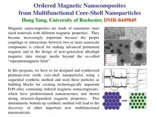
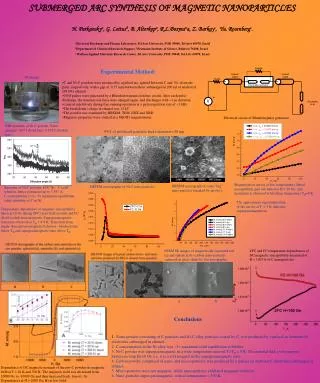
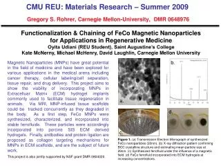
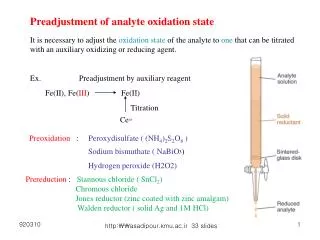
Process of Analyte Elimination by Chitosan-coated Magnetic Nanoparticles for CE-UV Analysis of Biochemically Active Compounds Meissam Noroozifar1, Zafar Iqbal2, Edward P.C. Lai2* 1University of Sistan & Baluchestan, Zahedan, Sistan & Baluchestan, Iran mnoroozifar@chem.usb.ac.ir 2Department of Chemistry, Carleton University, Ottawa, Ontario, Canada edward_lai@carleton.ca Abstract Results and Discussion Characterization of Particles MNPs, MNP@CH and MNPs@PDA were characterized by capillary electrophoresis with UV detection (at 200 nm) as shown in Fig. 4. Cationic chitosan-coated MNPs migrated faster than negatively-charged MNPs@PDA. Peaks appeared between 2.8 to 3.9 min (MNPs@CH) are due to the polydisperse particles. Fragmentation of long-chain high MW chitosan during coating would be a plausible explanation of this particles distribution. Whereas a single and fairly sharp peak at 4.29 min represents the monodisperse nature of MNPs@PDA. SEM image of the particles is presented in Fig. 4 that would support the monodispersity of MNPs@PDA. Chitosan-coated magnetic nanoparticles are a class of nanoparticles that has attracted much attention because of their advantageous characteristics, such as biochemical inertness, non-toxicity, bioconjugation ability, and potential for eliminating a variety of biochemically active compounds. Chitosan with two different molecular weights (low and high) have been successfully coated on magnetic nanoparticles. These chitosan-coated magnetic nanoparticles were tested as new sorbents for binding of biochemically active compounds under a wide range of sample matrix characteristics. Capillary electrophoresis (CE) was used to obtain results at the analytical scale that show that the nanoparticles can selectively interact with aromatic compounds. Their adsorption behavior under optimal experimental conditions has been modeled after isotherm equations. • FTIR spectroscopy • Different types of polydopamine (PDA) and chitosan (CH)-coated MNPs were first characterized by FTIR. As shown in Fig. 1, the characteristic peak of MNPs at 592 cm-1 appears weakened in all these spectra due to suppression by the coatings. Chitosan (CH)-coated MNPs are distinguishable from MNPs by characteristic peaks ranging from 900 to 1500 cm-1.Also, the appearance of two peaks around 2900 cm-1 indicates a thick coating of chitosan on the MNPs. In comparison, polydopamine (PDA)-coated MNPs were characterized by two peaks between 800 and 900 cm-1. Figure 1 Figure 4 Introduction Currently available treatment modalities for high-risk clinically localized prostate cancer have limited chances of achieving complete tumor elimination because of inadequate local or metastatic tumor eradication. Numerous phase I/II studies have been conducted to evaluate the safety and efficacy of neoadjuvant chemotherapy before prostatectomy. A multi-agent regimen of mitoxantrone (MTX) was reported to have antineoplastic activity as evidenced by reductions in prostate-specific antigen [1]. MTX is also a therapeutic compound approved by health authorities for the treatment of secondary progressive multiple sclerosis [2]. A novel capillary electrophoresis (CE) with chemiluminescence detection method was developed for the determination of MTX in commercial drug, human urine and plasma. It was based on sensitization of the reaction between potassium ferricyanide and luminol in sodium hydroxide medium, affording a detection limit of 1.0×10⁻⁸ M [3]. Continued study of novel agents in the neoadjuvant setting is warranted because this approach allows for the rapid identification of active agents and for molecular investigation into the mechanism of drug activity. Recently nontoxic magnetic nanoparticles (MNPs) have attracted a wide range of applications in medicine. Coating or functionalization of MNP surfaces can be better for drug delivery, targeted therapy, magnetic resonance imaging, transfection, and cell/protein/DNA separation [4]. MNPs have been coated with different materials (such as silica, octadecylsilane and polymers) to remove pollutants in water by different researchers [5,6,7]. Chitosan (CH) has strong affinity for binding certain endocrine disrupting compounds (EDCs) and pharmaceuticals that are biochemically active. An external magnetic field can then be applied to collect the loaded particles. After desorption of these compounds in a small volume of solvent, instrumental methods can be used to perform quantitative analysis. In the present work the chemical functionality of chitosan is investigated to examine its potential benefits to human health. Chitosan-Drug Binding Interactions Mitoxantrone in CH3CN/phosphate buffer (pH=8.5) and prednisolone in ethanol/phosphate buffer (pH=8.5) were next subjected to binding tests (Fig. 5). Based on CE-UV analysis results, MNPs@PDA-CH (high MW,70–150 kD) and MNPs@PDA were able to bind mitoxantrone very well. One plausible explanation of a lower % binding for prednisolone (pKa= 13.9) is its decreased hydrophobicity and aromaticity relative to mitoxantrone (pKa = 8.2 and 11.4) which has two hydrophobic phenyl groups. Chromatographic hydrophobicity measurements can be used in the summing of hydrophobicity values plus aromatic ring count [log DpH7.4 (or log P) + #Ar] to forecast property-based drug delivery by the particles studied in this work [11]. • Magnetic property Thepolydopamine-coated MNPs (or MNPs@PDA)and chitosan-coated MNPs (or MNPs@CH) were tested for their magnetic properties in air and aqueous suspension. Their strong attraction to the external magnet is illustrated in Fig. 2. Figure 2 Figure 5 Capillary electrophoresis analysis CE-UV analysis of organic compounds (200 µg.mL-1) was performed before and after in-vitro binding with MNPs@CH (10 mg.mL-1) in 20 mM Na2HPO4 (pH 8.5±0.2). A mixture including bisphenol A (BPA), metformin (MF), naphthalene acetic acid (NAA), phenformin (PF) and quinine sulphate (QS) was electrokinetically injected for 3 s at 17 kV and analyzed under 20 kV. Figure 3 shows that quinine sulfate and phenformin can be eliminated more efficiently than bisphenol A and metformin. Surprisingly, naphthalene acetic acid cannot be eliminated at all. One good example is that determination of metformin would be much less susceptible to interference by phenformin. Conclusions Before After Chitosan is a biodegradable and aqueous compatible polymer that has attracted great attention in the pharmaceutical development of nanoparticles for encapsulation of drugs and biological substances. Nontoxic polydopamine is known for its aqueous and biocompatibility. This work demonstrates the idea of in-vitroelimination of several analytes from a complex sample matrix by binding with polymer-coated magnetic nanoparticles before CE-UV analysis. The strong magnetic property of these particles indicates that MNPs@CHand MNPs@PDA could potentially be applied for delivering bound cancer drugs to a specific organ inside the patient. It would be necessary to determine the blood kinetics as dictated by their ionic charge states. More research will be needed to investigate the drug release properties of biodegradable chitosan and polydopamine. Experimental 2.1 Materials Chitosan with high molecular weight (CH(HM)) and low molecular weight (CH(LM)), bisphenol A (BPA), disodium hydrogen phosphate (Na2HPO4), dopamine (DA), iron(II) chloride tetrahydrate (FeCl2.4H2O), iron(III) chloride hexahydrate (FeCl3.6H2O), mesityl oxide (MO), metformin hydrochloride (MF.HCl), mitoxantrone (MTX), naphthalene acetic acid (NAA), phenformin (PF), prednisolone (PRED), pyrrole (Py), sodium dodecyl sulphate (SDS) and triclosan (TC) were all obtained from Sigma-Aldrich (Oakville, ON, Canada). HPLC-grade methanol (MeOH) and ethanol (EtOH) were purchased from Caledon (Georgetown, ON, Canada). 2.2 Preparation of coated magnetic nanoparticles MNPs (Fe3O4) were prepared using a method previously described by Wang et al. [8] through co-precipitation of Fe2+ and Fe3+ ions in solution by excess NaOH under sonication. MNPs were coated with polydopamine (PDA) using a procedure reported by Wenet al. [9] with sonication. Coating of MNPs or MNPs-PDA with chitosan (high and low molecular weights) was carried out as follows: 30 mL of Tris buffer (10 mM, pH = 9.2) including 50 mM SDS was sonicated in a 200 mL beaker for 10 min. Then, 2 ml of CH(HM) 1% m/m in 1% acetic acid (AA) or 20 ml CH(LM) 0.1% m/m in AA was added and the solution was sonicated for 10 min. Then, the solution was added to 1.0 g of MNPs in Tris buffer and the suspension was sonicated for 20 min. Finally the black coated MNPs were separated by a magnet, washed with DDW and EtOH (5 times each), and dried at room temperature for 24 hours. 2.3. CE-UV analysis A modular system was built in our laboratory for all capillary electrophoresis (CE) separations with UV detection at 200 nm. The background electrolyte (BGE) inside the capillary was 20 mM Na2HPO4 in DDW (pH 8.5), running under an applied voltage of 17–20 kV at ambient temperature. A Lambda 1010 UV detector (Bischoff, Leonberg, Germany) was set up at a wavelength of 200 nm to detect the migration of all analytes. The PeakSimple software (SRI Instruments, Torrance, California, USA) was used to acquire the detector output signal. The BGE in inlet and outlet vials was changed after every ten CE analyses to maintain high purity and constant level. Figure 3 Process of Analyte Elimination In-vitro elimination of organic compounds from a standard solution was investigated with three different MNPs (10 mg.mL-1) in 20 mM Na2HPO4 (pH 8.5±0.2). All % binding results are presented in Table 1 as the mean (± standard deviation) of triplicate measurements (n = 3). Phenformin binds up to 95% with MNPs@chitosan by hydrophobic interaction whereas metformin binds only up to 21%. The NAA results are very surprising because chitosan was commonly regarded to be cationic and able to interact with negatively charged molecules [10]. From their chemical point of view, chitosan represents a weak base. The antimicrobial efficiency of high MW chitosan was also explained by the binding of the cationic chitosan to the anionic molecules at the outer surface of the bacterial membrane. References [1] M. Garzotto, A. Myrthue, C.S.Higano and T.M. Beer, Urologic Oncology, 2006, 24, 254-259. [2] O. Neuhaus, B.C. Kieseier and H.P. Hartung, Pharmacology & Therapeutics, 2006, 109, 198-209. [3] S. Han and H. Wang, Journal of Chromatography B, 2010, 878(28), 2901-2904. [4] M.R. Shishehbore, A. Afkhami and H. Bagheri, Colloids & Surfaces B, 2011, 82, 316–324. [5] L. Chen, T. Wang and J. Tong, Trends in Analytical Chemistry, 2011, 30, 1095–1108. [6] M. Miah, Z. Iqbal and E.P.C. Lai, Analytical Methods, 2012, 4, 2866-2878. [7] Z. Iqbal, S. Alsudir, M. Miah and EPC Lai, Electrophoresis, 2011, 32, 1–7. [8] X. Wang, L. Wang, X. He, Y. Zhang and L. Chen, Talanta, 2009, 78, 327–332. [9] H.Z. Wen, H.L. Chun, C.G. Xiu, R.C. Fa, H.Y. Huang and R.W. Xiao, Journal of Materials Chemistry, 2010, 20, 880–883. [10] Y. Boonsongrit, A. Mitrevej and B.W. Mueller, European Journal of Pharmaceutics and Biopharmaceutics, 2006, 62, 267–274. [11] R.J. Young, D.V.S. Green, C.N. Luscombe and A.P. Hill, Drug Discovery Today, 2011, 16, 823-830. Table 1 Acknowledgement Financial support of the Natural Sciences and Engineering Research Council (NSERC) Canada is gratefully acknowledged.