Ch 4
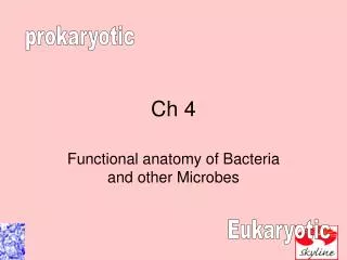
prokaryotic. Ch 4. Functional anatomy of Bacteria and other Microbes. Eukaryotic. Or The differences between Eukaryotic and Prokaryotic cells. Q&A. Penicillin was called a “miracle drug” because it doesn’t harm human cells. Why doesn’t it?.


Ch 4
E N D
Presentation Transcript
prokaryotic Ch 4 Functional anatomy of Bacteria and other Microbes Eukaryotic
Q&A Penicillin was called a “miracle drug” because it doesn’t harm human cells. Why doesn’t it?
Proks and euks are similar in chemical composition and reaction Proks lack membrane bound organelles Only Proks have peptidoglycan Euks have membrane bound organelles Euks have paired chromosomes Euks have histones
Nick sees the difference mainly in information and structural capacity • Proks lack membrane-enclosed organelles • Euks are like a 2mhz 100gb home computer • Proks are like a calculator • Human genome 4x10⁹ • E. coli 4x10⁶
The prokaryote • Unicellular • Multiply by binary fission • Differentiated by • Morphology • Chemical composition • Nutritional requirements • Biochemical activates • Sources of energy • Other tests
Size • 0.2 to 2um in diameter • 2-8um in length • In biological systems there are always exceptions these are general sizes.
Coccus Diplococci Streptococci Staphylococci Bacillus Spiral Other pleomorphic shapes Shape
Glycocalyx Capsule Slime layer Extracellular polysaccharide Function Toxicity Protect from phagocytosis Allow adherence Reduce water loss Collect nutrients Parts not seen
Used in movement Can present taxis Negative Positive Monotrichous Peritrichous Flagellar H protein acts as an antigen E.c O157:H7 Flagellin Flagella: long filamentous appendages with filament, hook and basal body
Flagella Arrangement Figure 4.7
Shorter and less complex than flagella Helps adhere to surfaces Used for sex and communication Fimbriae/pili
Major difference between eukaryotic and prok orgs. Surrounds plasma membrane provides protection Peptidoglycan Polymer of NAG NAM Short amino acid chain Production inhibited by antibiotics Prevents osmotic damage Damage to cw is almost always lethal except Cell wall
Peptidoglycan Polymer of disaccharide: N-acetylglucosamine (NAG) N-acetylmuramic acid (NAM) Figure 4.12
Peptidoglycan in Gram-Positive Bacteria Figure 4.13a
The Cell Wall Prevents osmotic lysis 4-7 Differentiate protoplast, spheroplast, and L form. Made of peptidoglycan (in bacteria) Linked by polypeptides Figure 4.6
Gram-Positive Bacterial Cell Wall Figure 4.13b
Gram-Negative Bacterial Cell Wall Figure 4.13c
Thick peptidoglycan Teichoic acids Gram-positiveCell Wall Gram-positiveCell Wall • Thin peptidoglycan • Outer membrane • Periplasmic space Figure 4.13b–c
Gram neg • Lipoprotein phospholipid outer membrane surrounding a thin peptidoglycan • Makes gram neg resistant to • Phagocytosis • Antibiotics • Chemical reactions • Enzymes (lysozyme) • Has lipid A endotoxin • O polysaccaride antigen O157:H7 E.c.
Gram-Negative Outer Membrane Figure 4.13c
The Gram Stain • Gram-Positive (b) Gram-Negative Table 4.1
The Gram Stain Mechanism Crystal violet-iodine crystals form in cell Gram-positive Alcohol dehydrates peptidoglycan CV-I crystals do not leave Gram-negative Alcohol dissolves outer membrane and leaves holes in peptidoglycan CV-I washes out
2-ring basal body Disrupted by lysozyme Penicillin sensitive Gram-PositiveCell Wall Gram-NegativeCell Wall • 4-ring basal body • Endotoxin • Tetracycline sensitive Figure 4.13b–c
Nontypical cell walls • Mycoplasma (acid fast) do not have ppt containing cell wall. • Archaea contain another chemical called pseudomurein
Atypical Cell Walls Acid-fast cell walls Like gram-positive Waxy lipid (mycolic acid) bound to peptidoglycan Mycobacterium Nocardia Figure 24.8
Atypical Cell Walls Mycoplasmas Lack cell walls Sterols in plasma membrane Archaea Wall-less or Walls of pseudomurein (lack NAM and D-amino acids)
Damage to the Cell Wall Lysozyme digests disaccharide in peptidoglycan Penicillin inhibits peptide bridges in peptidoglycan Protoplast is a wall-less cell Spheroplast is a wall-less gram-positive cell Protoplasts and spheroplasts are susceptible to osmotic lysis L forms are wall-less cells that swell into irregular shapes
Plasma membrane • Defines the living and nonliving parts of the cell • Everything on the inside is living • Everything on the outside is not living • Is selectively permeable • Workspace for enzymes of metabolic reactions
Plasma Membrane • Phospholipid bilayer • Peripheral proteins • Integral proteins • Transmembrane proteins Figure 4.14b
PM Workspace • Nutrient breakdown • Energy production • Photosynthesis • Afforded by mesosomes which are regular infoldings of the plasma membrane • Weaknesses: destroyed by actions of alcohols, detergents and polymyxins
Fluid Mosaic Model • Membrane is as viscous as olive oil. • Proteins move to function • Phospholipids rotate and move laterally Figure 4.14b
Plasma Membrane • Damage to the membrane by alcohols, quaternary ammonium (detergents) and polymyxin antibiotics causes leakage of cell contents.
Movement of Materials across Membranes Simple diffusion: Movement of a solute from an area of high concentration to an area of low concentration Figure 4.17a
Movement of Materials across Membranes Facilitated diffusion: Solute combines with a transporter protein in the membrane Figure 4.17b-c
Movement of Materials across Membranes ANIMATION Passive Transport: Special Types of Diffusion ANIMATION Passive Transport: Principles of Diffusion
Movement of Materials across Membranes Osmosis: The movement of water across a selectively permeable membrane from an area of high water to an area of lower water concentration Osmotic pressure: The pressure needed to stop the movement of water across the membrane Figure 4.18a