Download
1 / 82
820 likes | 1.34k Vues
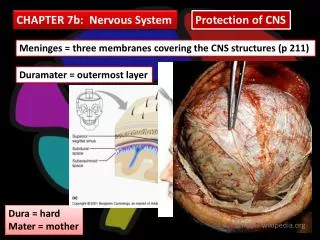
Chapter 11 Nervous System II. Meninges membranes surrounding CNS protect CNS three layers dura mater – outer, tough arachnoid mater - weblike pia mater – inner, delicate. Meninges of the Spinal Cord. Ventricles. interconnected cavities within cerebral hemispheres and brain stem

E N D
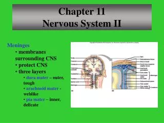
Chapter 11Nervous System II • Meninges • membranes surrounding CNS • protect CNS • three layers • dura mater – outer, tough • arachnoid mater - weblike • pia mater – inner, delicate
Ventricles • interconnected cavities • within cerebral hemispheres and brain stem • continuous with central canal of spinal cord • filled with cerebrospinal fluid (csf) • lateral ventricles • third ventricle • fourth ventricle • cerebral aqueduct
Cerebrospinal Fluid • secreted by choroid plexus • circulates in ventricles, central canal of spinal cord, and subarachnoid space • completely surrounds brain and spinal cord • clear liquid • nutritive and protective • helps maintain stable ion concentrations in CNS
Spinal Cord Structure • extends foramen magnum to 2nd lumbar vertebra
Spinal Cord Functions • center for spinal reflexes • conduit for nerve impulses to and from the brain
Reflex Arcs Reflexes – automatic, subconscious responses to stimuli
Knee-jerk Reflex • helps maintain posture
Withdrawal Reflex • protective
Crossed-Extensor Reflex • flexor muscles contract • flexor muscles on opposite side inhibited • extensor muscles on opposite side contract for balance
Tracts of the Spinal Cord • Ascending tracts conduct sensory impulses to the brain • Descending tracts conduct motor impulses from the brain to motor neurons reaching muscles and glands
Ascending Tracts • fasciculus cuneatus • lateral spinothalamic
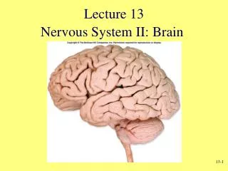
Brain • Functions • interprets sensations • determines perception • stores memory • reasoning • makes decisions • coordinates muscular movements • regulates visceral activities • determines personality • Major Parts • cerebrum • two cerebellar hemispheres • diencephalon • brain stem • cerebellum
Brain Development • Three Major Vesicles • Forebrain • Midbrain • Hindbrain • Forebrain (prosencephalon) • anterior portion (telencephalon) • cerebrum • basal ganglia • posterior portion (diencephalon) • thalamus • hypothalamus • posterior pituitary • pineal gland
Brain Development • Midbrain (mesencephalon) • midbrain • Hindbrain (rhombencephalon) • anterior portion (metencephalon) • cerebellum • pons • posterior portion (myelencephalon) • medulla oblongata
Structure of Cerebrum • corpus callosum • connects hemispheres • convolutions • bumps or gyri • sulci • grooves • longitudinal fissure • separates hemispheres • transverse fissure • separates cerebrum from cerebellum
Lobes of Cerebrum • Frontal • Parietal • Temporal • Occipital • Insula
Functions of Cerebrum • interpretation • initiating voluntary movements • storing memory • retrieving memory • reasoning • center for intelligence and personality
Functional Regions of Cerebral Cortex Cerebral Cortex – thin layer of gray matter that constitutes the outermost portion of cerebrum; contains 75% of all neurons in nervous system
Motor Areas • Primary Motor Areas • frontal lobes • control voluntary muscles • Broca’s Area • anterior to primary motor cortex • usually in one hemisphere • controls muscles needed for speech • Frontal Eye Field • above Broca’s area • controls voluntary movements of eyes and eyelids
Sensory Areas • Cutaneous Sensory Area • parietal lobe • interprets sensations on skin • Visual Area • occipital lobe • interprets vision • Auditory Area • temporal lobe • interprets hearing
Association Areas • regions of cortex that are not primary motor or primary sensory areas • widespread throughout the cerebral cortex • analyze and interpret sensory experiences • provide memory, reasoning, verbalization, judgment, emotions
Association Areas • Frontal Lobe Association Areas • concentrating • planning • problem solving • judging • Temporal Lobe Association Areas • remember visual scenes • remember music • remember complex patterns • Parietal Lobe Association Areas • understanding speech • using words to express thought • Occipital Lobe Association Areas • combine visual images with other sensory experiences
Hemisphere Dominance • In over 90% of population, left hemisphere is dominant • Nondominant hemisphere controls • nonverbal tasks • motor tasks • understanding and interpreting musical and visual patterns • provides emotional and intuitive thought processes • Dominant hemisphere controls • speech • writing • reading • verbal skills • analytical skills • computational skills
Memory • Short Term • working memory • closed circuit • circuit is stimulated over and over • when impulse flow stops, memory disappears • Long Term • changes structure and function of neurons • enhanced synaptic transmission
Basal Nuclei • masses of gray matter • deep within cerebral hemispheres • caudate nucleus, putamen, globus pallidus • produce dopamine • control certain muscular activities
Diencephalon • between cerebral hemispheres and brainstem • surrounds third ventricle • thalamus • hypothalamus • optic tracts • optic chiasm • infundibulum • posterior pituitary • mammillary bodies • pineal gland
Diencephalon • Thalamus • gateway for sensory impulses heading to cerebral cortex • receives all sensory impulses (except smell) • channels impulses to appropriate part of cerebral cortex for interpretation • Hypothalamus • maintains homeostasis by regulating visceral activities • links nervous and endocrine systems
Limbic System • Consists of • portions of frontal lobe • portions of temporal lobe • hypothalamus • thalamus • basal nuclei • other deep nuclei • Functions • controls emotions • produces feelings • interpret sensory impulses
Brain Stem • Three Parts • Midbrain • Pons • Medulla Oblongata
Midbrain • between diencephalon and pons • contains bundles of fibers that join lower parts of brainstem and spinal cord with higher part of brain • cerebral aqueduct • cerebral peduncles – bundles of nerve fibers • corpora quadrigemina – centers for visual and auditory reflexes
Pons • rounded bulge on underside of brainstem • between medulla oblongata and midbrain • helps regulate rate and depth of breathing • relays nerve impulses to and from medulla oblongata and cerebellum
Medulla Oblongata • enlarged continuation of spinal cord • conducts ascending and descending impulses between brain and spinal cord • contains cardiac, vasomotor, and respiratory control centers • contains various nonvital reflex control centers (coughing, sneezing, vomiting)
Reticular Formation • complex network of nerve fibers scattered throughout the brain stem • extends into the diencephalon • connects to centers of hypothalamus, basal nuclei, cerebellum, and cerebrum • filters incoming sensory information • arouses cerebral cortex into state of wakefulness
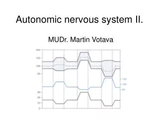
Types of Sleep • Slow Wave • person is tired • decreasing activity of reticular system • restful • dreamless • reduced blood pressure and respiratory rate • ranges from light to heavy • alternates with REM sleep • Rapid Eye Movement (REM) • some areas of brain active • heart and respiratory rates irregular • dreaming occurs
Cerebellum • inferior to occipital lobes • posterior to pons and medulla oblongata • two hemispheres • vermis connects hemispheres • cerebellar cortex – gray matter • arbor vitae – white matter • cerebellar peduncles – nerve fiber tracts • dentate nucleus – largest nucleus in cerebellum • integrates sensory information concerning position of body parts • coordinates skeletal muscle activity • maintains posture
Peripheral Nervous System • Cranial nerves arising from the brain • Somatic fibers connecting to the skin and skeletal muscles • Autonomic fibers connecting to viscera • Spinal nerves arising from the spinal cord • Somatic fibers connecting to the skin and skeletal muscles • Autonomic fibers connecting to viscera
Nerve Fiber Classification • Sensory Nerves – conduct impulses into CNS • Motor Nerves – conduct impulses to muscles or glands • Mixed Nerves – contain both sensory nerve fibers and motor nerve fibers; most nerves • General somatic afferent fibers • carry sensory impulses to CNS from skin and skeletal muscles • General somatic efferent fibers • carry motor impulses from CNS to skeletal muscles • General visceral efferent fibers • carry motor impulses away from CNS to smooth muscles and glands • General visceral afferent fibers • carry sensory impulses to CNS from blood vessels and internal organs
Nerve Fiber Classification • Special somatic efferent fibers • carry motor impulses from brain to muscles used in chewing, swallowing, speaking, and forming facial expressions • Special visceral afferent fibers • carry sensory impulses to brain from olfactory and taste receptors • Special somatic afferent fibers • carry sensory impulses to brain from receptors of sight, hearing, and equilibrium
Cranial Nerves I and II • Olfactory (I) • sensory • fibers transmit impulses associated with smell • Optic (II) • sensory • fibers transmit impulses associated with vision
Cranial Nerves III and IV • Oculomotor (III) • primarily motor • motor impulses to muscles that • raise eyelids • move the eyes • focus lens • adjust light entering eye • Trochlear (IV) • primarily motor • motor impulses to muscles that move the eyes
Cranial Nerve V • Trigeminal (V) • mixed • opthalmic division • sensory from surface of eyes, tear glands, scalp, forehead, and upper eyelids • maxillary division • sensory from upper teeth, upper gum, upper lip, palate, and skin of face • mandibular division • sensory from scalp, skin of jaw, lower teeth, lower gum, and lower lip • motor to muscles of mastication and muscles in floor of mouth
Cranial Nerves VI and VII • Abducens (VI) • primarily motor • motor impulses to muscles that move the eyes • Facial (VII) • mixed • sensory from taste receptors • motor to muscles of facial expression, tear glands, and salivary glands
Cranial Nerves VIII and IX • Glossopharyngeal (IX) • mixed • sensory from pharynx, tonsils, tongue, and carotid arteries • motor to salivary glands and muscles of pharynx • Vestibulocochlear (VIII) • sensory • sensory from equilibrium receptors of ear • sensory from hearing receptors