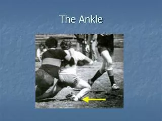
ANKLE
ANKLE. Divisions of the Foot. Hindfoot: Talus. Calcaneus. Midfoot: Navicular. Cuboid. Cuneiforms. Forefoot: Metatarsals. Phalanges. Ankle Joints and Ligaments. Superior and inferior tibiofibular joints: Superior: Synovial plane joint with a capsule. Inferior:

ANKLE
E N D
Presentation Transcript
Divisions of the Foot • Hindfoot: Talus. Calcaneus. • Midfoot: Navicular. Cuboid. Cuneiforms. • Forefoot: Metatarsals. Phalanges.
Ankle Joints and Ligaments • Superior and inferior tibiofibular joints: Superior: Synovial plane joint with a capsule. Inferior: Syndesmosis: Posterior tibiofibular ligament. Anterior tibiofibular ligament. Crural tibiofibular interosseous ligament.
Ankle Joints and Ligaments • Talotibial (talocrural): Most congruent joint in the body. Synovial hinge joint. Axis: Toe out stance (= normal tibial torsion). Pathological: external/internal tibial torsion.
Ankle Joints and Ligaments • Movements: Dorsiflexion = increased toe out. Plantarflexion = decreased toe out. Mostly occur in sagittal plane.
Ankle Joints and Ligaments • Talotibial (talocrural): Mortise and tenon joint. Mortise: Fibular malleolus. Tibial malleolus. Distal end of tibia. Tenon: Head of talus.
Ankle Joints and Ligaments • Talotibial (talocrural): Ligaments: Medial collateral = deltoid: Tibionavicular. Tibiocalcanean. Anterior tibiotalar. Posterior tibiotalar.
Ankle Joints and Ligaments • Talotibial (talocrural): Ligaments: Lateral collateral (weakest of the collaterals): Anterior talofibular. Posterior talofibular. Calcanefibular.
Ankle Joints and Ligaments • Talocalcaneal (subtalar): Very stable. Uniaxial, triplanar.
Ankle Joints and Ligaments • Talocalcaneal (subtalar): Three surfaces: Posterior: Concave facet on talus with convex facet on calcaneus. Anterior: Convex facets on body and neck of talus. Concave facets on calcaneus.
Ankle Joints and Ligaments • Talocalcaneal (subtalar): Tarsal tunnel: Nonsynovial but with ligament: Talocalcaneal ligament.
Ankle Joints and Ligaments • Talocalcaneal (subtalar): Movements: Inversion: Adduction (vertical axis). Supination (longitudinal axis). Plantarflexion (coronal axis). Eversion: Opposite inversion.
Ankle Joints and Ligaments • Talocalcaneal (subtalar): Ligaments: Interosseous talocalcaneal. Posterior and lateral talocalcaneal ligaments.
Ankle Joints and Ligaments • Talocalcaneonavicular: Talocalcaneal + talonavicular: Movements: Inversion. Eversion.
Ankle Joints and Ligaments • Talocalcaneonavicular: Ligaments: Calcaneonavicular (spring) ligament: From sustentacular tali to inferior navicular. Continuous medially with deltoid ligament. Continuous laterally with medial band of bifurcate ligament. Helps to maintain medial longitudinal arch.
Ankle Joints and Ligaments • Calcaneocuboid: Triplanar. Ligaments: Plantar calcaneocuboid (short plantar). Long plantar (most important).
Ankle Joints and Ligaments • Transverse tarsal: = talonavicular + calcaneocuboid. Adds to inversion-eversion range. Compensates forefoot for hindfoot eversion. Keeps distal foot inverted with lateral surface in contact with the ground while the hindfoot is everted.
Retinacula • Superior/inferior extensor retinacula: Localized thickenings of anterior crural fascia. Bind down tendons of: Tibialis anterior. Extensor hallucis longus. Extensor digitorum longus. Peroneus tertius.
Retinacula • Superior/inferior extensor retinacula: Covers: Deep peroneal nerve. Anterior tibial artery. Inferior retinaculum is “Y”-shaped.
Retinacula • Flexor retinaculum: Localized medial thickening of crural fascia. Binds down tendons of: Flexor hallucis longus. Flexor digitorum longus. Tibialis posterior. Covers: Tibial nerve. Posterior tibial artery.
Retinacula • Peroneal (fibular) retinaculum: Localized thickenings of lateral deep fascia. Binds down tendons of: Peroneus (fibularis) longus. Peroneus (fibularis) brevis.
Plantar Arches • Longitudinal: Medial. Lateral. • Pathologies: Pes cavus. Pes planus.
Plantar Arches • Support ligaments: Plantar calcaneonavicular (spring): Primary support for medial longitudinal arch. Long plantar: Primary support for lateral longitudinal arch.
Plantar Arches • Support ligaments: Plantar aponeurosis. Plantar calcaneocuboid (short plantar). Marginal abductors.