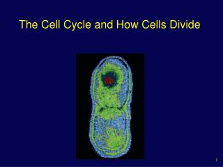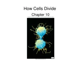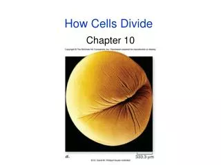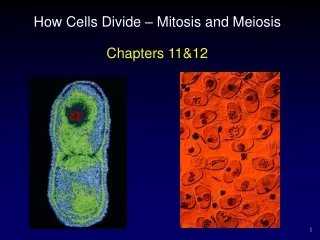
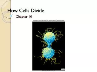
How Cells Divide
How Cells Divide. Chapter 10. Bacterial Cell Division. Bacteria divide by binary fission No sexual life cycle Reproduction is clonal Single, circular bacterial chromosome is replicated Replication begins at the origin of replication and proceeds bidirectionally to site of termination

How Cells Divide
E N D
Presentation Transcript
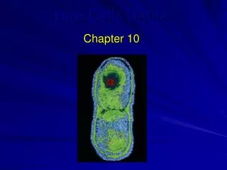
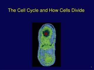
How Cells Divide Chapter 10
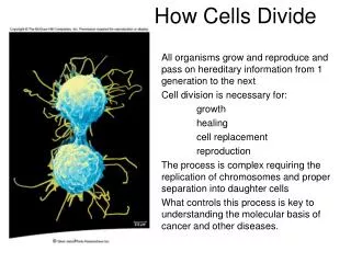
Bacterial Cell Division • Bacteria divide by binary fission • No sexual life cycle • Reproduction is clonal • Single, circular bacterial chromosome is replicated • Replication begins at the origin of replication and proceeds bidirectionally to site of termination • New chromosomes are partitioned to opposite ends of the cell • Septum forms to divide the cell into 2 cells (FtsZ protein)
Eukaryotic Chromosomes • Every species has a different number of chromosomes • Humans have 46 chromosomes in 23 nearly identical pairs • Additional/missing chromosomes usually fatal
Chromosomes • Composed of chromatin – complex of DNA and protein • DNA of a single chromosome is one long continuous double-stranded fiber • Typical human chromosome 140 million nucleotides long • In the nondividing nucleus • Heterochromatin – not expressed • Euchromatin – expressed
Structure • Nucleosome • Complex of DNA and histone proteins • Promote and guide coiling of DNA • DNA duplex coiled around 8 histone proteins every 200 nucleotides • Histones are positively charged and strongly attracted to negatively charged phosphate groups of DNA
Nucleosomes wrapped into higher order coils called solenoids • Leads to a fiber 30 nm in diameter • Usual state of nondividing (interphase) chromatin • During mitosis, chromatin in solenoid arranged around scaffold of protein to achieve maximum compaction • Radial looping aided by condensin proteins
Karyotype • Particular array of chromosomes in an individual organism • Arranged according to size, staining properties, location of centromere, etc. • Humans are diploid (2n) • 2 complete sets of chromosomes • 46 total chromosomes • Haploid (n) – 1 set of chromosomes • 23 in humans • Pair of chromosomes are homologous • Each one is a homologue
Replication • Prior to replication, each chromosome composed of a single DNA molecule • After replication, each chromosome composed of 2 identical DNA molecules • Held together by cohesin proteins • Visible as 2 strands held together as chromosome becomes more condensed • One chromosome composed of 2 sister chromatids
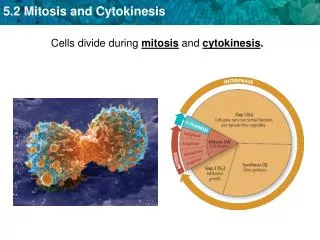
Eukaryotic Cell Cycle • G1 (gap phase 1) • Primary growth phase, longest phase • S (synthesis) • Replication of DNA • G2 (gap phase 2) • Organelles replicate, microtubules organize • M (mitosis) • Subdivided into 5 phases • C (cytokinesis) • Separation of 2 new cells Interphase
Duration • Time it takes to complete a cell cycle varies greatly • Fruit fly embryos = 8 minutes • Mature cells take longer to grow • Typical mammalian cell takes 24 hours • Liver cell takes more than a year • Growth occurs during G1, G2, and S phases • M phase takes only about an hour • Most variation in length of G1 • Resting phase G0 – cells spend more or less time here
Interphase • G1, S, and G2 phases • G1 – cells undergo major portion of growth • S – replicate DNA • G2 – chromosomes coil more tightly using motor proteins; centrioles replicate; tubulin synthesis • Centromere – point of constriction • Kinetochore – attachment site for microtubules • Each sister chromatid has a centromere • Chromatids stay attached at centromere by cohesin • Replaced by condensin in metazoans
M phase Mitosis is divided into 5 phases: 1. Prophase 2. Prometaphase 3. Metaphase 4. Anaphase 5. Telophase
Prophase • Individual condensed chromosomes first become visible with the light microscope • Condensation continues throughout prophase • Spindle apparatus assembles • 2 centrioles move to opposite poles forming spindle apparatus (no centrioles in plants) • Asters – radial array of microtubules in animals (not plants) • Nuclear envelope breaks down
Prometaphase • Transition occurs after disassembly of nuclear envelope • Microtubule attachment • 2nd group grows from poles and attaches to kinetochores • Each sister chromatid connected to opposite poles • Chromosomes begin to move to center of cell – congression • Assembly and disassembly of microtubules • Motor proteins at kinetochores
Metaphase • Alignment of chromosomes along metaphase plate • Not an actual structure • Future axis of cell division
Anaphase • Begins when centromeres split • Key event is removal of cohesin proteins from all chromosomes • Sister chromatids pulled to opposite poles • 2 forms of movements • Anaphase A – kinetochores pulled toward poles • Anaphase B – poles move apart
Telophase • Spindle apparatus disassembles • Nuclear envelope forms around each set of sister chromatids • Now called chromosomes • Chromosomes begin to uncoil • Nucleolus reappears in each new nucleus
Cytokinesis • Cleavage of the cell into equal halves • Animal cells – constriction of actin filaments produces a cleavage furrow • Plant cells – cell plate forms between the nuclei • Fungi and some protists – nuclear membrane does not dissolve; mitosis occurs within the nucleus; division of the nucleus occurs with cytokinesis
Please note that due to differing operating systems, some animations will not appear until the presentation is viewed in Presentation Mode (Slide Show view). You may see blank slides in the “Normal” or “Slide Sorter” views. All animations will appear after viewing in Presentation Mode and playing each animation. Most animations will require the latest version of the Flash Player, which is available at http://get.adobe.com/flashplayer.
Control of the Cell Cycle • Current view integrates 2 concepts • Cell cycle has two irreversible points • Replication of genetic material • Separation of the sister chromatids • Cell cycle can be put on hold at specific points called checkpoints • Process is checked for accuracy and can be halted if there are errors • Allows cell to respond to internal and external signals
3 Checkpoints • G1/S checkpoint • Cell “decides” to divide • Primary point for external signal influence • G2/M checkpoint • Cell makes a commitment to mitosis • Assesses success of DNA replication • Late metaphase (spindle) checkpoint • Cell ensures that all chromosomes are attached to the spindle
Cyclin-dependent kinases (Cdks) • Enzymes that phosphorylate proteins • Primary mechanism of cell cycle control • Cdks partner with different cyclins at different points in the cell cycle • For many years, a common view was that cyclins drove the cell cycle – that is, the periodic synthesis and destruction of cyclins acted as a clock • Now clear that Cdk itself is also controlled by phosphorylation
Cdk – cyclin complex • Also called mitosis-promoting factor (MPF) • Activity of Cdk is also controlled by the pattern of phosphorylation • Phosphorylation at one site (red) inactivates Cdk • Phosphorylation at another site (green) activates Cdk
MPF • Once thought that MPF was controlled solely by the level of the M phase-specific cyclins • Although M phase cyclin is necessary for MPF function, activity is controlled by inhibitory phosphorylation of the kinase component, Cdc2 • Damage to DNA acts through a complex pathway to tip the balance toward the inhibitory phosphorylation of MPF
Anaphase-promoting complex (APC) • Also called cyclosome (APC/C) • At the spindle checkpoint, presence of all chromosomes at the metaphase plate and the tension on the microtubules between opposite poles are both important • Function of the APC/C is to trigger anaphase itself • Marks securin for destruction; no inhibition of separase; separase destroys cohesin
Control in multicellular eukaryotes • Multiple Cdks control the cycle as opposed to the single Cdk in yeasts • Animal cells respond to a greater variety of external signals than do yeasts, which primarily respond to signals necessary for mating • More complex controls allow the integration of more input into control of the cycle
Growth factors • Act by triggering intracellular signaling systems • Platelet-derived growth factor (PDGF) one of the first growth factors to be identified • PDGF receptor is a receptor tyrosine kinase (RTK) that initiates a MAP kinase cascade to stimulate cell division • Growth factors can override cellular controls that otherwise inhibit cell division
Cancer • Unrestrained, uncontrolled growth of cells • Failure of cell cycle control • Two kinds of genes can disturb the cell cycle when they are mutated • Tumor-suppressor genes • Proto-oncogenes
Tumor-suppressor genes • p53 plays a key role in G1 checkpoint • p53 protein monitors integrity of DNA • If DNA damaged, cell division halted and repair enzymes stimulated • If DNA damage is irreparable, p53 directs cell to kill itself • Prevent the development of mutated cells containing mutations • p53 is absent or damaged in many cancerous cells
Proto-oncogenes • Normal cellular genes that become oncogenes when mutated • Oncogenes can cause cancer • Some encode receptors for growth factors • If receptor is mutated in “on”, cell no longer depends on growth factors • Some encode signal transduction proteins • Only one copy of a proto-oncogene needs to undergo this mutation for uncontrolled division to take place
Tumor-suppressor genes • p53 gene and many others • Both copies of a tumor-suppressor gene must lose function for the cancerous phenotype to develop • First tumor-suppressor identified was the retinoblastoma susceptibility gene (Rb) • Predisposes individuals for a rare form of cancer that affects the retina of the eye
Inheriting a single mutant copy of Rb means the individual has only one “good” copy left • During the hundreds of thousands of divisions that occur to produce the retina, any error that damages the remaining good copy leads to a cancerous cell • Single cancerous cell in the retina then leads to the formation of a retinoblastoma tumor • Rb protein integrates signals from growth factors • Role to bind important regulatory proteins and prevent stimulation of cyclin or Cdk production
Please note that due to differing operating systems, some animations will not appear until the presentation is viewed in Presentation Mode (Slide Show view). You may see blank slides in the “Normal” or “Slide Sorter” views. All animations will appear after viewing in Presentation Mode and playing each animation. Most animations will require the latest version of the Flash Player, which is available at http://get.adobe.com/flashplayer.