Download
1 / 24
240 likes | 384 Vues
Structure of the cardiovascular System. What you do!. Copy the text with a white background. Those with a pink background are for information only, and notes on these will be found in your monograph. Components of the CVS.

E N D
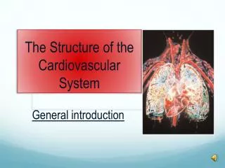
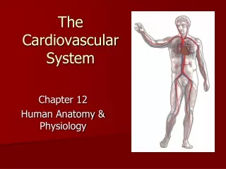
Structure of the cardiovascular System
What you do! • Copy the text with a white background. Those with a pink background are for information only, and notes on these will be found in your monograph
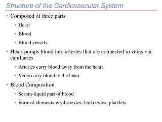
Components of the CVS • The CVS consists of a double pump (the heart) and a complex system of blood vessels to transport oxygen carrying blood around the body.
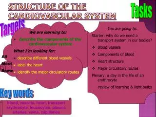
Blood Vessels • There are 3 types of blood vessel: • Arteries (and arterioles) • Capillaries • Veins (and venules)
Arteries Arteries (and arterioles) carry blood away from the heart. The largest arteries e.g. the Aorta, have thick elastic walls which can stretch to accommodate the surge of blood after each contraction of the heart. Arteries branch many times, forming smaller and smaller vessels, the smallest of which are arterioles. Contraction of the smooth muscle lining the walls of the arterioles allows them to open or close to varying degrees to adjust blood flow to different parts of the body e.g. during vasodilation and vasoconstriction.
Veins Veins (and venules) carry blood back to the heart. Blood flows out of the capillaries into the smallest of the veins – venules – which in turn reunite to form larger veins. The walls of veins are thinner than those of arteries as blood pressure is far lower as it travels through veins. Consequently, veins have valves to prevent the back flow of blood. Oxygenated blood leaves the heart from the left ventricle via the aorta, moves through arteries to arterioles to capillaries to venules and returns to the right atrium by way of veins.
Capillaries Capillaries are tiny vessels where the exchange of substances with the tissue occurs. They also connect the arterioles to the venules. Their walls are only one cell thick, allowing nutrients and waste to diffuse through with ease. Capillaries form extensive branching networks (capillary beds) throughout the body tissues, but only certain beds are open at any one time. This allows the ‘shunting’ of the blood from one region to another. Arteriole Venule Capillaries (capillary bed)
The flow of blood through the capillaries is controlled by the contraction of a ring of muscle called a sphincter. This is important as the body does not contain sufficient blood for all vessels to be filled with blood at one time. This is understandable when we consider that that an individual could have between 25,000 to 60,000 miles of capillaries!
The CVS consists of 2 distinct circuits • The pulmonary circuit carrying deoxygenated blood from RV to lungs • The systemic circuit carrying oxygenated blood from LV to the aorta and then the rest of the body
The cardiac cycle • Each heartbeat is called a cardiac cycle and consists of the following • Atria contract simultaneously • Ventricles contract simultaneously • All chambers relax • Lasts about 0.8 secs (0.3systole,0.5 diastole)
Two phases of the cardiac cycle • Systole: contraction of the heart (Atrial first, the ventricular) • Diastole: relaxation of the heart
Cardiac Output • Cardiac Output is the volume of blood pumped by each ventricle per minute and is the function of two factors: • Heart rate (beats per minute) • Stroke volume (the volume of blood ejected by each ventricle during each contraction) • CO = HR x SV
At rest: HR = 72bpm SV = 70ml i.e. CO = 72 x 70 = 5040 ml/min = 5 litres/min Cardiac Output varies between individuals and depends on their physical fitness and level of activity. For example, the heart of a highly trained athlete can pump 30-35 l/min while most non-athletes can only achieve a max of 20 litres.
Some typical values for cardiac output at varying levels of activity
As work load increases, HR increases to a maximal value of about 180 – 200 bpm (220 minus age), while SV increases proportionally less (70-150ml). The increase in cardiac output with exercise is achieved principally by increasing the heart rate.
Blood Pressure The force exerted by the blood against the walls of the blood vessels is known as blood pressure
Measurement of BP Both systolic and diastolic BP can be measured by an inflatible instrument called a sphygmomanometer which is wrapped around the upper arm.
BP is measured in millimeteres of mercury (mm Hg) • Normal reading for a healthy adult is about 120/70 mm Hg (SBP/DBP) • As we age, BP rises due to atherosclerosis. A 65 yr old man may have a BP of 140/90