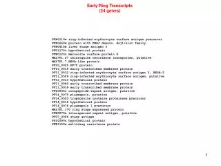
The Erythrocyte
The Erythrocyte. Henry O. Ogedegbe, PhD., BB(ASCP), C(ASCP)SC, CC(NRCC) Department of EHMCS . Learning Objectives. Upon completion of the materials in this chapter, the student will be able to: List sites of erythrocyte production and destruction


The Erythrocyte
E N D
Presentation Transcript
The Erythrocyte Henry O. Ogedegbe, PhD., BB(ASCP), C(ASCP)SC, CC(NRCC) Department of EHMCS
Learning Objectives • Upon completion of the materials in this chapter, the student will be able to: • List sites of erythrocyte production and destruction • Draw a flow diagram of the major erythrocyte morphologic maturation process • Describe classic morphologic features of each erythrocyte maturation stage • List each major erythrocyte component and its primary functions • Compare and contrast the destruction of normal and severely damaged cells
Normal Erythrocyte Production, Physiology and Destruction • The primary function of the erythron is to deliver oxygen to the tissues and carbon dioxide from the tissues • To function effectively, the body needs approximately 309 X 109 circulating erythrocytes per kilogram of body weight • The production and destruction of erythrocytes is kept in balance • The erythrocytes live for 120 days after which they are removed from the circulation by the cells of the RE system • They are promptly replaced to maintain the balance
Normal Erythrocyte Production, Physiology and Destruction • Origin: • The erythrocytes originate from a pluripoten stem cell called the colony forming unit-S (CFU-S) • During development, the cells become specialized and synthesize proteins needed for function and survival • As the cell matures the morphology changes. • There are stages in the maturation process • These stages take place in the bone marrow and may be differentiated by light microscopy • The burst-forming unit erythroid is the earliest erythroid committed cell
Normal Erythrocyte Production, Physiology and Destruction • The BFU-E is closely related to the CFU-S and matures into the CFU-E • The terms BFU-E and CFU-E are laboratory descriptions of growth patterns in culture media • Only a few BFU-E are present in the bone marrow and therefore are difficult to identify morphologically • They are different from but similar to small to medium sized lymphocytes • Increased numbers of BFU-E have been demonstrated in some anemias
Normal Erythrocyte Production, Physiology and Destruction • Production Sites: • The main sites of adult blood cell production include the vertebrae, pelvis, ribs, sternum, skull and the long bones • Radioactive imaging have been used to study the site of blood cell production • Iron is the radioisotope of choice because it mimics ingested iron which is bound to transferrin in the blood • Studies can be carried out in cases of anemia or after radiation treatment to evaluate production sites
Erythrocyte Maturation • Erythropoietin and other growth factors stimulate BFU-E and CFU-E to differentiate to the rubriblast stage • The rubriblast is the first precursor that can be recognized by light microscopy • The rubriblast gives rise to sixteen mature erythrocytes through four cell divisions which take about 72 hours • Changes take place in the rubriblast as it matures and differentiates from a primitive nucleated cell to a mature non-nucleated cell
Erythrocyte Maturation • Ultrastructure: • Organelles are present in the early erythrocyte which are necessary for the synthesis of hemoglobin, and proteins • As the proteins accumulate, the number of organelles gradually diminish • Differentiation of the maturing erythrocyte results in alterations in morphology and membrane properties • This results from reorganization of membrane skeletal protein network • An important component of the network is protein 4.1 • Protein 4.1 serves as a critical link between the cytoskeleton and the lipid bilayer
Erythrocyte Maturation • Nucleus: • The nucleus is very important in the earliest stages of red cell development • It is the site of DNA and RNA synthesis and thus critically involved in the red cell development and maturation • Chromatin contains genetic material and is composed of DNA, histones, and other proteins • The chromatin is finely dispersed and appears condensed or granular • The more condensed heterochromatin are inactive
Erythrocyte Maturation • The heterochromatin take on a basophilic color (dark blue) with basic dyes • The active euchromatin does not stain with basic dyes • As the cell matures chromatin becomes more dense and metabolic and synthetic activities start to decline • Finally the nucleus becomes inactive and it is extruded from the cell • Nucleoli are present in the rubriblast and they contain RNA, proteins and DNA • Nucleoli are involved with synthesis of ribosomal RNA
Erythrocyte Maturation • Cytoplasm: • Ribosomes and polyribosomes are present in the early erythrocyte precursor • They are the sites of globin and other protein synthesis • Polyribosomes probably synthesize different proteins from those synthesized by the ribosome • Ribosomes give the cytoplasm of early precursors a deep, dark blue color • As hemoglobin is formed, the number of ribosomes diminish and the blue color is replaced by a reddish pink color
Erythrocyte Maturation • Golgi apparatus is also present in the early precursor and is located near the nucleus • The Golgi is involved with protein modification within the cell • The mitochondria is also visible under electron microscope as rod shaped organelles • They are involved with aerobic generation of energy for the maturing cell and insertion of ferrous iron into protoporphyrin IX during heme synthesis • Iron is present in the cytoplasm as ferritin and hemosiderin
Erythrocyte Maturation • Maturation Stages: • Six morphological stages of erythrocyte maturation may be identified from a bone marrow sample with Wright stain • Normal maturation is dependent on intake of proper nutrients and vitamins such as folate, vitamin B12 and iron • Nomenclature: • There are three nomenclatures used to describe the six stages • Rubri (proposed by the ASCP) • Erythroblast (proposed by Paul Ehrlich) • Normoblast (normal precursor)
Erythrocyte Maturation • General Guidelines: • The ASCP published the following general guidelines for identification of erythroid precursors: • Progressive decrease in size and the degree of cytoplasmic basophilia (blue color) as the cell matures • Nuclei are round or oval in the blast stage become round thereafter • Gradual increase in coarseness and condensation of the chromatin, ranging from fine in the early stages to pyknotic in the stage just before nuclear extrusion
Erythrocyte Maturation • Rubriblast (Pronormoblast): • This is the earliest erythrocyte precursor identifiable by light microscopy in a Wright stained bone marrow prep • Cell size ranges from 12 to 25 m • The nuclear:cytoplasmic ratio is high • Nucleus usually occupies more than 80% of cell • The cytoplasm stains basophilic due to high RNA content • The Golgi may be visible near the nucleus usually pale
Erythrocyte Maturation • Prorubricyte (Basophilic normoblast): • Slightly smaller (12 – 17 m) than a rubriblast • Nucleus usually occupies 75% of the cell • The cytoplasm is basophilic and the Golgi is usually visible near the nucleus • The nucleus is round and its chromatin is dark violet and coarser and more clumped • The nucleoli is absent and helps in the identification • The prorubricyte usually divides two times giving rise to four rubricytes
Erythrocyte Maturation • Rubricyte (Polychromatophilic normoblast: • It is usually smaller than the prorubricyte (12-15 m) • Has a round nucleus that may be eccentric • The nucleus is smaller and the cytoplasm becomes more prominent • There is a spectrum of blue color due to synthesis of hemoglobin • The RNA and hemoglobin give the cytoplasm a blue gray violet color called polychromasia or polychromatophilia • The cell may be confused with a lymphocyte
Erythrocyte Maturation • Metarubricyte (Orthochromic normoblast): • This is the last nucleated erythrocyte stage • It is slightly smaller than the rubricyte (8-12m) • The cytoplasm is polychromatophilic and more pinkish than that of the rubricyte • The nuclear chromatin is dense, coarse and clumped • The nucleus is degenerated and pyknotic • The nucleus is extruded from the cell at this stage • Sometimes nucleus is not completely extruded resulting in Howell-Jolly body
Erythrocyte Maturation • Reticulocyte (diffusely basophilic erythrocyte): • The reticulocyte is slightly larger than the mature erythrocyte • The cytoplasm still contains small amounts of RNA which produces varying amount of polychromasia • The reticulocytes are retained in the bone marrow for 2 to 3 days before release into the marrow sinisoids • The mechanism of release is unknown • The retic contain Golgi apparatus remnant and residual mitochondria which permit continued aerobic metabolism
Erythrocyte Maturation • The retic also contain RNA which may be stained supravitally with methylene blue or brilliant cresyl blue • The RNA precipitates and the retics can then be counted and the reticulocyte production index determined • Mature Erythrocyte: • The mature erythrocyte is approximately 7.2 m in diameter • It is a biconcave disc and hence referred to as a discocyte • In a Wright stain, a central pale area is revealed which fades gradually into the reddish pink cytoplasm
Structure and Physiology of the Mature Erythrocyte • The mature erythrocyte lacks a nucleus or organelles • Components necessary for function and survival are present • The cell has a specialized membrane that allows for O2 and CO2 transport and for survival for 120 days • Various factors contribute to erythrocyte membrane and hemoglobin maintenance • A source of energy is required • Membrane shape and deformability are needed
Structure and Physiology of the Mature Erythrocyte • Shape and Deformability: • The erythrocyte is a biconcave disc which facilitates O2 and CO2 transport by maximizing ratio of surface area to volume • Allows the cell to be flexible and deformable • This allows the cell to adjust to small vessels in the microvasculature and still maintain a constant surface area • A less deformable cell would be subjected to fragmentation
Structure and Physiology of the Mature Erythrocyte • Membrane Composition and Structure: • The composition of the membrane allows the cell to • Separate the intracellular fluid environment of the cytoplasm from the extracellular fluid environment • Selectively pass nutrients and ions into and out of cell • Deform when required • The membrane is composed of lipids and proteins in approximately equal proportions by weight • The difference in the lipids and proteins in the cytoplasmic side and the plasma side allow for selective movement of molecules in and out of the cell
Structure and Physiology of the Mature Erythrocyte • Lipids: • Phospholipids and unesterified cholesterol predominate in the lipid fraction • The phospholipids form the bilayer and the hydrophilic polar heads of the phospholipids are oriented toward the aqueous environments • The hydrophobic tails of the phospholipids are oriented to the interior of the bilayer • The phospholipids are fluid and the fatty acid tails move freely • Cholesterol plays an important role in maintaining surface area
Structure and Physiology of the Mature Erythrocyte • Protein: • Proteins are bound to lipids throughout the membrane • The proteins are either peripheral proteins or integral proteins • The peripheral proteins are present on the inner portion of the membrane nearest the cytoplasm • The integral proteins are in contact with both the inner and the outer surface of the membrane • The integral proteins act as receptors for ions and molecules needed in the cell such as transferrin and EPO
Structure and Physiology of the Mature Erythrocyte • The peripheral proteins include the and spectrin also called band 1 and 2 and actin • The proteins form the cytoskeleton of the cell and regulate membrane shape and deformability • Their linkage is mediated by protein 4.1 • The principal integral proteins are glycoproteins designated glycophorin A and band 3 • They span the lipid bilayer. • Band 3 is an inorganic anion transport channel • Integral proteins contain sialic acid which gives erythrocytes a negative charge
Structure and Physiology of the Mature Erythrocyte • The negativity between cells called zeta potential cause cells to repel one another as they move through the circulation • Membrane proteins facilitate movement of substrates and cofactors in and out cell • Examples include the Na+, K+ - ATPase and Ca2+, Mg2+ - ATPase • Calcium is involved in regulation of and stabilization of membrane phospholipid structure • High intracellular concentration of calcium, cause cell deformability
Structure and Physiology of the Mature Erythrocyte • Energy Metabolism: • The cell requires energy for cell metabolism and to preserve the membrane integrity • Various enzymatic reactions in the cell require energy • Energy is required to reduce proteins and maintain hemoglobin in its reduced state for proper functioning • Two site prone to oxidation are the iron atom in the heme ring and the sulfhydryl groups on the globin molecule • Oxidation of the normal ferrous state to the ferric state results in methemoglobin which does not deliver oxygen
Structure and Physiology of the Mature Erythrocyte • Normally 1% to 3% of oxygen is oxidized to methemoglobin • Oxidation of sulfhydryl groups causes hemoglobin precipitation (Heinz body formation) • Sources of Energy: • The Embden-Meyerhof Pathway (EMP): • This is an anaerobic process for energy generation through glucose catabolism to lactate • About 90% to 95% of glucose used by the cells is metabolized by the EMP
Structure and Physiology of the Mature Erythrocyte • ATP is generated during the glycolysis of glucose to lactate • ATP is needed to maintain membrane shape and deformability • Through phosphorylation of spectrin and calcium chelation • Provide energy for active transport of cations • And to modulate the amount of 2,3 DPG generated • There is a net yield of two ATP molecules per molecule of glucose catabolized • 2,3 DPG is formed from the Rapoport-Luebering shunt • Helps modulate O2 transport in the cell
Structure and Physiology of the Mature Erythrocyte • Hexose Monophosphate Shunt and Glutathione Reduction pathway: • Also called the pentose phosphate pathway is an aerobic method of erythrocyte glycolysis • Processes about 10% of erythrocyte glucose • Purpose is to provide reducing potential by generating reduced nicotinamide adenine dinucleotide phosphate (NADPH) • It is an oxidative pathway
Erythrocyte Destruction • As the red cell ages, changes occur that make it susceptible to destruction • Alteration in the membrane integrity takes place • Loss of sialic acid and lipids, decreased ATP and increased Calcium have been implicated in the aging process • At 120 days the erythrocytes are recognized as abnormal and are removed by phagocytic cell in the RES • As the cell ages it is depleted of glucose and their surface area decreases • The spleen recognizes abnormalities in the cell and sequester it for removal
Regulation of Erythropoiesis • A balance between production and destruction keeps the the erythrocyte number constant • Production of erythron requires a normal functioning competent bone marrow • Adequate levels of EPO, growth factors and nutrients such as iron, folate, vitamin B12 • Between 3 X 10⁹ and 8.5 X 10⁹ erythrocytes are produced daily • Cytokines or growth factors play a important part in the process
Regulation of Erythropoiesis • Erythropoietin Production and Regulation: • EPO is a glycoprotein hormone with a molecular weight of 34,000 • It is an erythroid growth factor • It is produced in the kidney • It is regulated by renal O2 tension which when decreased induces expression of the EPO gene and the release of EPO • Prostaglandins help regulate EPO production
Regulation of Erythropoiesis • Growth Factors: • Many other hormones and cytokines secreted by various cells have been found to stimulate erythropoiesis • These cytokines have been identified in cell cultures and they include • EPO • Insulin • Growth hormone • Steroid hormone • Nonandrogenic thyroid hormone • IL-1, IL-4, IL-6, IL-7, IL-11, IL-12, • G-CSF • Macrophage inflammatory protein (MIP) and steel factor (SF)