Arrhythmia
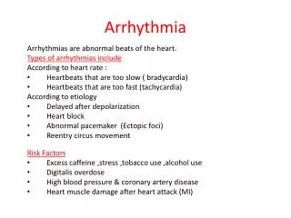
Arrhythmia. Rhythm refers to the regularity or spacing of the ECG waves. Any variation from the normal rhythm and sequence of excitation of the heart is termed an arrhythmia. Or simply Abnormal rhythm of heart is called arrhythmia. Causes of Arrhythmias.


Arrhythmia
E N D
Presentation Transcript
Arrhythmia • Rhythm refers to the regularity or spacing of the ECG waves. Any variation from the normal rhythm and sequence of excitation of the heart is termed an arrhythmia. Or simply • Abnormal rhythm of heart is called arrhythmia.
Causes of Arrhythmias • Abnormal rhythmicity of the pacemaker • Shift of the pacemaker from the sinus node to another place in the heart • Blocks at different points in the spread of the impulse through the heart • Abnormal pathways of impulse transmission through the heart • Spontaneous generation of abnormal impulses in almost any part of the heart
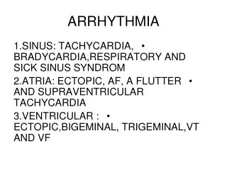
Tachycardia • The term “tachycardia” means fast heart rateusually defined in an adult person asfaster than 100 beats per minute.
Bradycardia • The term “bradycardia” means a slow heart rate, usually defined as fewer than 60beats per minute.
Sinus Arrhythmia • Under normal conditions the heart quickens during inspiration and slows down during expiration due to change in the autonomic nerve supply to the heart
Abnormal Rhythms That Result from Block of Heart Signals Within the Intracardiac Conduction Pathways • SinoatrialBlock • Atrioventricular Block
Sinoatrial Block • The impulse from the sinus node is blocked before it enters the atrial muscle. • The ventricles pick up a new rhythm the impulse usually originating spontaneously in the A-V node, so that the rate of the ventricular QRS-T complex is slowed but not otherwise altered
Atrioventricular Block • The only means by which impulses ordinarily can pass from the atria into the ventricles is through the A-V bundle, also known as the bundle of His.
Causes • Ischemia of the A-V node or A-V bundle fibers • Compression of the A-V bundle by scar tissue or by calcified portions of the heart can depress or block conduction from the atria to the ventricles. • Inflammation of the A-V node or A-V bundle • Extreme stimulation of the heart by the vagusnerves
Incomplete Atrioventricular Heart Block • First-Degree Heart Block The usual lapse of time between beginning of the P wave and beginning of the QRS complex is about 0.16 second when the heart is beating at a normal rate. This so called P-R interval usually decreases in length with faster heart beat and increases with slower heartbeat. When the P-R interval increases to greater than 0.20 second the P-R interval is said to be prolonged and the patient is said to have first degree incomplete heart block.
Second-degree Heart Block • When conduction through the A-V bundle is slowed enough to increase the P-R interval to 0.25 to 0.45 second, the action potential sometimes is strong enough to pass through the bundle into the ventricles and sometimes is not strong enough. In this case there will be an atrial P wave but no QRS-T complex and there are dropped beats of the ventricles. This condition is called second degree heart block.
Third degree Heart Block • When the condition causing poor conduction in the A-V node or A-V bundle becomes severe, complete block of the impulse from the atria into the ventricles occurs. In this case the ventricles spontaneously establish their own signal usually originating in the A-V node or A-V bundle. • The P waves become dissociated from the QRS-T complexes. The ventricles have escaped from control by the atria and they are beating at their own rate controlled most often by rhythmical signals generated in the A-V node or A-V bundle.
Stokes-Adams Syndrome • In some patients with A-V block the total block comes and goes. That is impulses are conducted from the atria into the ventricles for a period of time and then suddenly impulses are not conducted. When impulses are not conducted there are periodic fainting spells in these patients which is called Stokes-Adams Syndrome.
Ventricular Escape • Each time A-V conduction ceases the ventricles often do not start their own beating until after a delay of 5 to 30 seconds. This results from the phenomenon called overdrive suppression. This means that ventricular excitability is at first in a suppressed state because the ventricles have been driven by the atria at a rate greater than their natural rate of rhythm. After few seconds some part of the Purkinje system beyond the block usually in the distal part of the A-V node or in the A-V bundle begins discharging rhythmically at a rate of 15 to 40 times per minute and acting as the pacemaker of the ventricles.
Premature Contractions • A premature contraction is a contraction of the heart before the time that normal contraction would have been expected.This condition is also called extrasystole, premature beat or ectopic beat.
Causes of Premature Contractions • Most premature contractions result from ectopic foci in the heart which emit abnormal impulses at odd times during the normal cardiac rhythm.
Causes of Ectopic Focus • Ischemia • Small calcified plaques at different points in the heart which press against the adjacent cardiac muscle so that some of the fibers are irritated • Toxic irritation of the A-V node, Purkinje system, or myocardium caused by drugs, nicotine, caffeine. • Mechanical initiation of premature contractions is also frequent during cardiac catheterization
Compensatory Pause • The interval between the premature contraction and the next succeeding contraction is slightly prolonged which is called a compensatory pause.
Cause • The premature contraction originated in the atrium some distance from the sinus node and the impulse had to travel through a considerable amount of atrial muscle before it discharged the sinus node. The sinus node discharged late in the premature cycle and this made the succeeding sinus node discharge also late in appearing.
The term paroxysmal means that the heart rate becomes rapid in paroxysms, with the paroxysm beginning suddenly and lasting for a few seconds, a few minutes, a few hours or much longer. Then the paroxysm usually ends as suddenly as it began with the pacemaker of the heart instantly shifting back to the sinus node.
Paroxysmal tachycardia often results from an aberrant rhythm that involves the A-V node. This usually causes almost normal QRS-T complexes but totally missing or obscured P waves. • Atrial or A-V nodal paroxysmal tachycardia both of which are called supraventricular tachycardias. It usually occurs in young otherwise healthy people and they generally grow out of the predisposition to tachycardia after adolescence.
Ventricular Fibrillation • Ventricular fibrillation is a very serious rhythmic abnormality in which the ventricular musculature exhibits uncoordinated chaotic contractions. Multiple impulses travel erratically in all directions around the ventricles. • Ventricular fibrillation results from cardiac impulses that have gone berserk within the ventricular muscle mass, stimulating first one portion of the ventricular muscle, then another portion, then another and eventually feeding back onto itself to re-excite the same ventricular muscle over and over—never stopping.
Many small portions of the ventricular muscle will be contracting at the same time while equally as many other portions will be relaxing. There is never a coordinate contraction of all the ventricular muscle at once which is required for a pumping cycle of the heart.
After fibrillation begins unconsciousness occurs within 4 to 5 seconds for lack of blood flow to the brain and irretrievable death of tissues begins to occur throughout the body within a few minutes
CAUSES • Sudden electrical shock of the heart • Ischemia of the heart muscle • Ischemia of specialized conducting system
Re-entry-Circus Movements as the Basis for Ventricular Fibrillation
A long pathway typically occurs in dilated hearts. • Decreased rate of conduction frequently results from (a) blockage of the Purkinje system (b) ischemia of the muscle (c) high blood potassium levels (d) many other factors • A shortened refractory period commonly occurs in response to various drugs, such as epinephrine or after repetitive electrical stimulation. Thus in many cardiac disturbances re-entry can cause abnormal patterns of cardiac contraction or abnormal cardiac rhythms.
Treatment • Electroshock Defibrillation of the Ventricles • Hand Pumping of the Heart (Cardiopulmonary Resuscitation) as an aid to defibrillation
A moderate alternating-current voltage applied directly to the ventricles throws the ventricles into fibrillation, a strong high voltage alternating electrical current passed through the ventricles for a fraction of a second can stop fibrillation by throwing all the ventricular muscle to refractoriness simultaneously. The current penetrates most of the fibers of the ventricles at the same time, thus stimulating essentially all parts of the ventricles simultaneously and causing them all to become refractory. All action potentials stop and the heart remains silent for 3 to 5 seconds after which it begins to beat again. • Fibrillation can usually be stopped using 110 volts of 60-cycle alternating current applied for 0.1second or 1000 volts of direct current applied for a few thousandths of a second.
Atrial Fibrillation • Atrial fibrillation is characterized by rapid, irregular, uncoordinated depolarizationsof the atria with no definite P waves. • The common cause of atrial fibrillation is atrial enlargement resulting from heart valve lesions that prevent the atria from emptying adequately into the ventricles or from ventricular failure with excess damming of blood in the atria. The dilated atrial walls provide ideal conditions for a long conductive pathway as well as slow conduction both of which predispose to atrial fibrillation.
For the same reasons that the ventricles will not pump blood during ventricular fibrillation neither do the atria pump blood in atrial fibrillation. • Blood flows passively through the atria into the ventricles and the efficiency of ventricular pumping is decreased only 20 to 30 per cent.
Atrial Flutter • Atrial flutter is characterized by a rapid but regular sequence of atrial depolarizations at rates between 200 and 380 beats per minute. The ventricles rarely keep pace with the racing atria. Because the conducting tissue’s refractory period is longer than that of the atrial muscle, the AV node is unable to respond to every impulse that converges on it from the atria. • The electrical signal travels as a single large wave always in one direction around and around the atrial muscle mass. • Maybe only one out of every two or three atrial impulses successfully passes through the AV node to the ventricles. Such a situation is referred to as a 2:1 or 3:1 rhythm.
Because one side of the atria is contracting while the other side is relaxing the amount of blood pumped by the atria is slight
Atrial tachycardia-220/min • Atrial Flutter-200-350/min • Atrial Fibrillation-300-500/min