Download

1 / 31
320 likes | 383 Vues
Learn about epithelial tissues in Histology Lab 1, including classification, processing techniques, and common stains. Lab procedures and scoring breakdown provided. Reserve lab facilities for practical study.

E N D
Histology Lab 1 Introduction and Epithelium Jun Zhou(周俊), Ph.D & M.D School of Medicine,Zhejiang University
Note: • Please come lab on time. • Find your seat according your lab number.
Your final score is composed of four parts 1) Attendance and picture drawing:10% 2) Quiz: 15% (each quiz 5%) 3) Final lab test: 25% 4) Final written examination: 50% How to pass this course: Final score>60 and Final written exam>50
Lab facilities: • 1 Light electron microscope with computer • 1 case of slides • 1 atlas • http://m-learning.zju.edu.cn 1.Students share these facilities 2.Do not take them out the laboratory with you. 3.Put them where they belong to when lab is over. 4.Do not left any rubbish after you. 5.No food in the lab. 6.Lecture and lab slides online: m-learning.zju.edu.cn 2015 Histology and Embryology
Contents of Lab 1: • Introduction • Epithelium • 1)Simple squamous epi (small intestine,stomach) • 2)Simple cuboidal epi.(thyroid) • 3)Simple columnar epi.(small intestine)(drawing) • 4)Pseudostratified ciliated columnar epi.(trachea) • 5)Stratified squamous epi.(esophagus) • 6) Transitional epi.(bladder) • 3.Lab report (content of lab , drawing)
Tissue Processing 1.Fixation: 10% Formalin 2.Dehydration:graded concentrated ethyl alcohol 3.Clearing:xylene,infiltrate with paraffin solvent. Loss of fat due to ‘clearing’
5.Sectioning Tissue Processing 4.Embedding
Tissue Processing 6.Staining
Common Stains: Hematoxylin & Eosin Ross, 5th ed, fig 1.1
Common Stains Hematoxylin and eosin ("H&E") Wright’s stain
Relative Sizes in Histology Standard Units of Measure
Relative Sizes in Histology Standard Units of Measure nanometer: 10-9 meter micrometer: 10-6 meter
Changes in apparent shape, due to orientation of sections Ross, 5th ed, fig 1.11
Changes in apparent shape, due to orientation of sections Netter/Ciba Junqueira fig 1-30
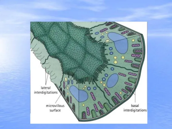
Epithelium Epithelium • No blood vessles, nourishment is by diffusion • Epithelium is innervated • polarization Trachea
Classification of Epithelia • Number of cells • 1. Simple • 2. Pseudostratified • 3. Stratified • Shape of surface cells • 1. Squamous • 2. Cuboidal • 3. Columnar • 4. Transitional
Stomach (NO 25): mesothelium HE ×400 Simple squamous epi.
Thyroid: HE × 400 Simple cuboidal epi.
Small intestine (NO 1): villus HE × 400 (Drawing ) Simple columnar epi., Goblet cells, striated border (microvilli)
Trachea Pseudostratified ciliated columnar epi
Trachea: HE × 400 Pseudostratified ciliated columnar epi.
Esophagus (No.2) Stratified squamous epi.
distended contracted Transitional epithelium Bladder
Bladder: HE × 100 Transitional epi.
Bladder: HE × 400 Transitional epi.