INTRODUCTION TO HISTOLOGY
701 likes | 2.38k Vues
INTRODUCTION TO HISTOLOGY. By Dr. A.K. Akinloye Department of Veterinary Anatomy University of Agriculture Abeokuta. What is Histology?. The term histology, is derived from the Greek histos , meaning tissue (web) and logia , knowledge

INTRODUCTION TO HISTOLOGY
E N D
Presentation Transcript
INTRODUCTION TO HISTOLOGY By Dr. A.K. Akinloye Department of Veterinary Anatomy University of Agriculture Abeokuta
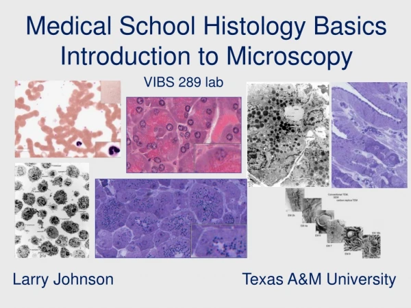
What is Histology? • The term histology, is derived from the Greek histos, meaning tissue (web) and logia, knowledge • It is, in the strict sense, the knowledge, or science, of tissues whether they are of plants or animals • is the study of the microscopic anatomy of cells and tissues of plants and animals • It is performed by examining a thin slice (section) of tissue under a light or electron microscope
What is Veterinary Histology? • Is the science that focuses on the detailed morphology of tissues of domestic animals with the aid of microscope and correlates specific structures with function
What is Veterinary Microanatomy? • Involves the examination and architectural description of the microscopic anatomy of normal cells of the body and all their contents and products
HISTORY Robert Hooke was the first person to observe cells in 1665. He looked at thin slices of cork under a very simple microscope. The cork appeared as little boxes which he called cells In 1883 Mathias Schleiden and Theodor Schwann proposed that all plants and animals were composed of cells which were the basic building blocks of life In 1855 Rudolf Virchow stated that new cells arise from the division of pre-existing cells and that chemical reactions needed for life occurred inside the cell All this work led to the formation of the cell theory
LEVELS OF ORGANIZATION • Cells • Tissues • Organs • Organ Systems • Organism
What is a Cell? • Is defined as the smallest basic structure of higher organisms capable of independent existence
What is a Tissue? • Is a group of cells of similar function and origin that form functional units
What is an Organ? • An organ represent an even greater measure of complexity and is composed of various tissues
What is an Organ System? • At an even higher level of organization: An organ system composed of several organs (such as the gastrointestinal system, respiratory system, cardiovascular system, endocrine system)
What is an Organism? • An organism can be seen to be formed of different levels of organization, with increasing levels of complexity and each of which plays important roles in the physiological homeostasis of the body
Histological Terms • The photographing of stained cells is called Histography or Photomicrography • Histopathology is the microscopic study of diseased tissue • The trained scientists who perform the preparation of histological sections are Histotechnicians, Histology Technicians (HT), Histology Technologists (HTL), Medical Scientists, Medical Laboratory Technicians or Biomedical scientists • Their field of study is called Histotechnology
Source of Tissue • Histological examination of tissues starts with surgery, biopsy or autopsy (or necropsy, in the case of animal tissues). • Biopsy is an examination of tissue taken from a living body • Autopsy is an examination of post-mortem tissue • Necropsy is an examination of tissue taken from dead animal
Technical Procedure • Fixation • The tissues are mechanically and biochemically stabilized in a fixative. The most common fixative is neutral buffered formalin (10% formaldehyde in phosphate buffered saline (PBS))
Technical Procedure • Embedding • Sectioning • Staining
Technical Procedure • Processing • The most common technique is wax processing. The samples are immersed in multiple baths of progressively more concentrated ethanol to dehydrate the tissue, followed by a clearing agent such as, xylene or Histoclear, and finally hot molten paraffin wax (impregnation). During this 12 to 16 hour process, paraffin wax will replace the xylene:
Staining • Routine staining is done to give contrast to the tissue being examined, as without staining it is very difficult to see differences in cell morphology • Hematoxylin and eosin (abbreviated H&E) are the most commonly used stains in histology and histopathology. Hematoxylin colours nuclei blue, eosin colours the cytoplasm pink • To see the tissue under a microscope, the sections are stained with one or more pigments
Special Staining • Other compounds used to colour tissue sections include: • safranin • oil red o • congo red • fast green FCF • silver salts • numerous natural and artificial dyes
Histochemistry • refers to the science of using chemical reactions between laboratory chemicals and components within tissue. A commonly performed histochemical technique is the Perls Prussian blue reaction, used to demonstrate iron deposits in diseases like Hemochromatosis
Immunohistochemistry • Recently, antibodies are used to specifically visualize proteins, carbohydrates and lipids: this is called Immunohistochemistry • Other advanced techniques include in situ hybridization to identify specific DNA or RNA molecules, and confocal microscopy
Alternative techniques • Cryosection - the tissue is frozen and cut using a cryostat • Tissue staining methods are similar to those of wax sections • Plastic embedding is commonly used in the preparation of material for electron microscopy. • Tissues are embedded in epoxy resin. • Very thin sections (less than 0.1 micrometers) are cut using diamond or glass knives. • The sections are stained with electron dense stains (uranium and lead) so that they can be seen with the Electron Microscope
Histological Classification of Animal Tissues • There are four basic types of tissues: • Epithelial tissue • Muscle tissue • Connective tissue • Nervous tissue
Epithelial tissue • A sheet of aggregated cells of a similar type tightly adhered to each other, constitutes the external and internal surfaces of the body • Epithelium: the lining of glands, bowel, skin and some organs like the liver, lung, kidney, • Endothelium: the lining of blood and lymphatic vessels, • Mesothelium: the lining of pleural, and pericardial spaces,
CONNECTIVE TISSUE • Connective tissue is responsible for providing structural support for the tissues and organs of the body. • This mechanical function is important in maintaining the form of the body, organs and tissues. • Connective tissue is composed of: • cells • extracellular matrix.
MUSCLE TISSUE • Muscle tissue is characterized by its well-developed properties of contraction • Muscle is responsible for the movements of the body and the various parts of the body • Muscle develops from embryonic mesoderm Muscle is classified into 3 categories according to morphology and physiological function: • Skeletal Muscle • Cardiac Muscle • Smooth Muscle
NERVOUS TISSUE • Nervous tissue consists of two groups of cell types: • Nerve cells (Neurons) • Neuroglia. • nervous tissue is derived from embryonic neuroectoderm • The nervous system is divided anatomically into: • Central Nervous System (CNS), consisting of the brain and spinal cord. • Peripheral Nervous System (PNS), consisting of nerve fibers, aggregates of nerve cells and glia and ganglia.