Download
1 / 41
1.28k likes | 4.94k Vues
Introduction to Oral Histology. Dr Firas Alsoleihat, BDS, PhD Department of Conservative Dentistry. Why do we study Oral Histology and Biology?. To understand the structure and function of oral tissues. To understand the development of oral tissues. To understand the general oral physiology.

E N D
Introduction to Oral Histology Dr Firas Alsoleihat, BDS, PhD Department of Conservative Dentistry
Why do we study Oral Histology and Biology? • To understand the structure and function of oral tissues. • To understand the development of oral tissues. • To understand the general oral physiology. • To understand oral diseases.
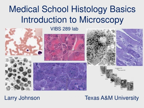
How Can We Study Oral Histology and Biology?? • Gross Anatomy • Physical properties • Chemical composition • Histological sections: • Hard tissues • Ground sections • Decalcified Sections • Soft Tissues
Oral Structures • Teeth: enamel, dentine, cementum and pulp. • Periodontium: gingiva, alveolar bone, periodontal ligament and root cementum. • Jaw bones. • Tempromandibular joint. • Oral mucosa. • Sub-mucosa: Blood vessels, Nerves. • Salivary glands.
Oral Structures • Hard tissues • Soft tissues
Oral Structures • Hard tissues: • Enamel • Dentine • Cementum • Alveolar bone • Jaw bones. • Tempromandibular joint.
Oral Structures • Soft tissues: • Pulp. • Gingiva • Periodontal ligament • Oral mucosa. • Sub-mucosa: Blood vessels, Nerves. • Salivary glands
Tooth structure - A tooth has a crown and root(s) with a pulp chamber and root canal(s). -Enamel, dentine, pulp tissue and cementum make up a tooth.
Enamel • Most highly mineralized tissue in the body • 96% inorganic material. • Non-vital, insensitive, cannot be regenerated.
Dentine • Dentine forms the bulk of the tooth. • It is rigid but elastic therefore ideal to support enamel. • It has a tubular structure • It is a vital, sensitive. • Capable of repair. • Formed throughout life.
Dentine • Primary dentine • secondary dentine • Tertiary dentine .
The Pulp • The pulp forms, nourishes, innervates and repairs dentine. • Soft connective tissue contained within the pulp chamber and the root canals.
Tooth Supporting Structures Teeth are supported by the perodontium which consists of: 1- The gingiva 2- Root cementum 3- Periodontal ligament 4- Alveolar bone
The Gingiva • The gingiva has 2 main regions: • the attached gingiva • the free gingiva.
Cementum • Thin layer of calcified tissue covering the dentine of the root. • Cementum varies in thickness at different levels of the root • thicker at the root apex and inter-radicular areas.
Cementum • The prime function of cemntum is to give attachment of the tooth to collagen fibres of the periodontal ligament. • Cementum can be repaired and regenerated
Periodontal Ligament • Dense fibrous connective tissue that attaches the tooth to the alveolar bone. • The periodontal space varies according to the functional state of teeth. • It is responsible for the functional position of the tooth; eruption, support (recovery after heavy loads) and drift.
Alveolar Bone • The part of the maxilla and mandible that supports the teeth. • Bone remodelling according to the functional demands . • Alveolar bone requires functional stimuli to maintain mass, otherwise it atrophies. • Outer and inner cortical plates • Individual tooth sockets are separated by inter-dental septa.
Jaw Bones • The maxilla and the mandible form the upper and lower jaw bones. • Histology of bone: compact and spongy bone.
Tempromandibular Joint • The TMJ is the synovial articulation between the mandible and the cranium.
Oral Mucosa • The oral mucosa represents the lining of the oral cavity • It consists of oral epithelium and an underlying connective tissue (lamina propria), and the basement membrane in between.
Oral Mucosa • The oral epithelium is poly-stratified squamous epithelium.The epithelial cells (keratinocytes) are arranged in layers.
Oral Mucosa • Depending on the location and function of the epithelium, the oral mucosae are classified into: • masticatory mucosa • lining mucosa • specialized mucosa.
Masticatory Oral Mucosa • Masticatory mucosa covers parts of the hard palate and the gingiva. • the epithelium is keratinised to withstand masticatory forces.
Lining Oral Mucosa • Lining mucosa covers the lips, cheeks, alveolar mucosa, soft palate, ventral surface of the tongue and the floor of the mouth.
Specialized Oral Mucosa Specialized oral mucosae include: 1- The mucosa covering the dorsal surface of the tongue. 2- The lingual tonsils. 3- The gingival attachment to teeth. 4- The vermillion border of the lip
Specialized Oral Mucosa The dorsal surface of the tongue • Characterized by the presence of lingual papillae. • Some of the papillae possess a mechanical function, while others have a sensory function (taste buds)
Specialized Oral Mucosa Lingual Tonsils • Lingual tonsils are situated at the posterior third of the tongue, at the lateral borders.
Specialized Oral Mucosa Gingival attachment to the tooth • Characterized by the presence of double basement membranes, one facing the connective tissue and the other facing the tooth.
Oral Sub-Mucosa • Oral sub-mucosa is a layer of loose fatty or glandular connective tissue. • This layer contains major blood vessels and nerves supplying the mucosa and separating it from underlying bones and muscles..
Salivary Glands • Three pairs of major salivary glands and minor salivary glands. • Major salivary glands: • Parotid • Submandibular • Sublingual
Conclusion It is important to understand the normal structure, development and function of oral structures, in order to understand the nature of pathologies faced in clinical practice.
http://blackboard.ju.edu.jo/ User name: danatomy_std Password: danatomy_std Click on course documents.
Reference Book • Berkovitz B., Holland G. and Moxham B.: Oral Anatomy, Histology and Embryology, 3rd or 4th edition, Edinburgh, 2005, Mosby. • Nanci A.:Ten Cate's Oral Histology: Development, Structure, and Function, 6th Edition, 2003, Mosby. Antonio, PhD • Bhaskar S.: Orban’s Oral Histology and Embryology, 11th edition, 1991, Mosby.


![[PDF] DOWNLOAD Notes on Oral Histology, Oral Physiology and Dental Anatomy](https://cdn7.slideserve.com/12532774/slide1-dt.jpg)