Histology: Introduction & Epithelial Tissue
821 likes | 1.95k Vues
Histology: Introduction & Epithelial Tissue. J. Matthew Velkey matt.velkey@duke.edu 452A Davison. Resources. For the STUDENT:. For the TEAM:. Textbook: Junqueira’s Basic Histology, 12 th ed. (each student should have a copy) Atlas: Color Atlas of Histology,

Histology: Introduction & Epithelial Tissue
E N D
Presentation Transcript
Histology:Introduction & Epithelial Tissue J. Matthew Velkey matt.velkey@duke.edu 452A Davison
Resources For the STUDENT: For the TEAM: Textbook: Junqueira’s Basic Histology, 12th ed. (each student should have a copy) Atlas: Color Atlas of Histology, 5th ed. by Garter & Hiatt (a copy is provided for each team to use during “lab” sessions) Online laboratory guide: http://www.duke.edu/web/histology/DPT.html When possible, lectures will be recorded and there may be notes for some lectures, but still NOT a substitute for reading the text. Completing assigned reading prior to class is essential for sessions where a READINESS ASSESSMENT is scheduled
Overall Objectives To understand: • How cells and tissues are arranged in the normal organ system of the body, and • How these cells and tissues are specialized to perform the function(s) most effectively. The knowledge gained will hopefully provide a cellular and ultrastructural “framework” for all of the other topics (anatomy, physiology, biochemistry, etc.) that you’ll learn this year. Histology is also, of course, a FUNDAMENTAL part of PATHOLOGY.
Correlate Structure and Function
HISTOLOGYThe study of cells and tissues, a.k.a. micro-anatomy
Tissue Preparation for Light Microscopy • Stabilize cellular structures by chemical fixation. • Dehydrate and infiltrate tissues with paraffin or plastic. • Embed fixed tissues in paraffin or plastic blocks. • Cut into thin slices of 3-10 micrometer thick; collect sections on slides. • Re-hydrate and stain with Hematoxylin (a basic dye): Stains basophilic structures (e.g. nucleic acids) blue/purple. • Counter-stain with Eosin (an acidic dye): Stains acidophilic or “eosinophilic” structures (e.g. proteins, membranes) red/pink. “H & E” staining is routine, but other dyes and staining techniques may be used to visualize other structures.
Light Microscopy 1. ILLUMINATION SOURCE 2. CONDENSER LENS 3. SPECIMEN STAGE 4. OBJECTIVE LENS 5. PROJECTION (OCULAR) LENS 6. OBSERVER • YIELDS A 2-DIMENSIONAL IMAGE CAPABLE OF 0.2 m RESOLUTION. • CELLULAR FEATURES ARE STAINED DIFFERENTIALLY BASED PRIMARILY UPON CHEMICAL PROPERTIES.
Light Microscopy Eosin (red): stains (+) charged structures, e.g. membranes and proteins Hematoxylin (blue): stains (-) charged structures, e.g. nucleic acids (DNA and RNA) and sulfated proteoglycans
Electron Microscopy WHY? The resolution of a microscope (the smallest distance two points can still be seen as separate points) is directly proportional to the wavelength of the radiation used. SOLUTION: • Tissues are fixed with glutaraldehyde (cross-links proteins) and osmium tetraoxide (cross-links lipids); OsO4 is also an electron-dense “stain” • Dehydrate and infiltrate tissues w/ plastic. • Embed and block fixed tissues in plastic. • Cut into ultra-thin slices (50 nanometers thick); collect sections on slides. • Stain sections with heavy metal salts (lead citrate and uranyl acetate) that bind nucleic acids & proteins. 6. Visualize in TEM; heavy metal “stains” block electrons to create contrast Radiation Wavelength Resolution Visible light 700-400 nm 0.2 µm Electrons 0.004 nm 0.1 nm PROBLEM: how to view tissue with a 30kV electron beam
Transmission Electron Microscopy 1. ILLUMINATION SOURCE (generates electron beam) 2. CONDENSER LENS 3. SPECIMEN STAGE 4. OBJECTIVE LENS 5. PROJECTION LENS 6. FLUORESCENT VIEW SCREEN 7. VIEWING WINDOW & OBSERVER • YIELDS A 2-DIMENSIONAL IMAGE CAPABLE OF 0.2 nm RESOLUTION. • CELLULAR FEATURES ARE STAINED WITH ELECTRON-DENSE, HEAVY METAL STAINS YIELDING ONLY A BLACK AND WHITE IMAGE
A cell’s form reflects its function e.g., plasma cells: highly specialized for the secretion of antibodies (proteins).
ORGANS are comprised of different TISSUES: Epithelial tissue Connective tissue Muscle Tissue Nerve Tissue e.g., the intestine Submucosa (connective tissue) Mucosa (epithelium + ct) Lumen Mesentery (ct + epithelium) Myenteric plexus (nerve) MuscularisExterna(smooth muscle)
[ Fr. Tissu, woven ; L. texo, to weave ] Tissues A tissue is an organized aggregation of cells or groups of cells that function in a coordinated manner to perform one or more specific functions. Tissues combine to form larger functional units, called ORGANS. Thus, the tissues are the basic functional units responsible for maintaining body functions.
BASIC TISSUES Epithelium Connective tissue Muscle Nervous tissue [Blood]
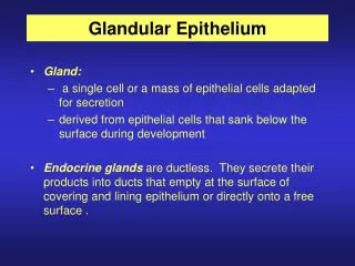
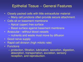
An epithelium is a cohesive sheet of cells that: 1. Covers the external surfaces and lines the internal surfaces of the body. • Barrier: Protection (by withstanding wear and tear, from hydration and dehydration) Selective absorption: (Control the movement of substances between the outside environment and the internal compartments, or between compartments in the body.) • Transport (ions, O2 and C02) • Secretion (secretory cells) 2. Forms endocrine and exocrine secretory glands. duct secretory portion Junquueira & Carneiro 10th Ed. P. 82
Epitheliallining cells ofSkin Intestine Multiple layers of cells with different shapes Single layer of tall (columnar) cells
Epithelial cells: 1. Form avascular sheets that differ in number of cell layers, shape of the cells and structural specializations of the free (apical) cell surface, depending on the tissue function(s). • Are capable of renewal and regeneration. non-specialized epithelium - all cells specialized epithelium - stem cells 3. Are structurally and functionally polarized: Have apical, lateral and basal domains. 4. Are held together by several basolateral specializations, known as the intercellular junctions, and bind to the underlying connective tissue via the basement membrane (LM) or basal lamina (EM).
Classification of Epithelium columnar (Respiratory)
Simple squamous epithelium: endothelium and mesothelium (non-specialized: renewal via mitosis)
Simple Cuboidal Epithelium kidney tubules (“non-specialized:” renewal via mitosis)
Simple Columnar Epithelium Gut mucosa (“specialized:” renewal via stem cells)
Simple columnar epitheliumlining the gut lumen lumen Two layers of smooth muscle on the wall
Stratified Squamous Epithelium non-keratinized keratinized Kierszenbaum pg 5
Stratified Squamous Epithelium Non-keratinized Keratinized Lines esophagus, oral cavity, vagina… Lines thick and thin skin
Transitional Epithelium(urothelium) Kierszenbaum pg 6
Transitional Epithelium(urothelium)Lines the urinary tract, ureter, bladder and urethra Cells on the surface are often dome (umbrella) shaped and some cells reveal two nuclei.
Pseudostratified Epithelium Kierszenbaum pg 6
Epithelial cells: 1. Form avascular sheets that differ in number of cell layers, shape of the cells and structural specializations of the free (apical) cell surface, depending on the tissue function(s). • Are capable of renewal and regeneration. non-specialized epithelium - all cells specialized epithelium - stem cells 3. Are structurally and functionally polarized: Have apical, lateral and basal domains. 4. Are held together by several basolateral specializations, known as the intercellular junctions, and bind to the underlying connective tissue via the basement membrane (LM) or basal lamina (EM).
Apical Cell Surface Specializations – 1Microvilli – aka “brush border” or “striated border” G G G: goblet cell
Microvilli(Core of actin filaments) NON-motile; serve to increase surface area
Apical Surface Specializations-2Cilia on Pseudostratified Columnar Epithelium with Goblet cells (Respiratory Epithelium) (from K. Verhey)
Cilia (Apical Cell Surface Specializations – 2)core of microtubules in 9+2 arrangement (axoneme) cilia Goblet cells Basal bodies Respiratory epithelium
9 + 2 (Axoneme)
Dynein is responsible for the sliding. Alberts et al., P. 648
Two types of apical cell surface specializations:Microvilli and cilia Microvilli Cilia
Epithelial cells: 1. Form avascular sheets that differ in number of cell layers, shape of the cells and structural specializations of the free (apical) cell surface, depending on the tissue function(s). • Are capable of renewal and regeneration. non-specialized epithelium - all cells specialized epithelium - stem cells 3. Are structurally and functionally polarized: Have apical, lateral and basal domains. 4. Are held together by several basolateral specializations, known as the intercellular junctions, and bind to the underlying connective tissue via the basement membrane (LM) or basal lamina (EM).
Basolateral Specializations Structures that hold the cells together and attach the epithelium to the underlying connective tissue. Intercellular junctions can only be observed at the electron microscope level and NOT at the light microscope level. Basement membrane (basal lamina)
Macula adherens (desmosomes) and Intermediate Filaments Desmosomes are NOT visible at the light microscope level.
Desmosomes and Intermediate Filaments Desmosomes serve as: 1. Spot attachment sites for adjacent cell membranes. 2. Anchoring sites for intermediate filaments. (from K. Verhey) Alberts et al., p. 802
Hemidesmosomes function to anchor epithelial cells to their basement membrane. Basement membrane
Loss of desmosome functions cause Blistering Skin Disorders Pemphigus: Separation of epidermal cells from each other (acantholysis) caused by loss of desmosome functions. Bullous pemphigoid: Separation of epidermis from the dermis due to blistering in the basement membrane caused by loss of anchoring filaments and hemidesmosomes.