Download
1 / 23
230 likes | 346 Vues
Learn about RNA structure, transcription steps (initiation, elongation, termination), mRNA processing (capping, splicing), and role in protein synthesis. Discover the genetic code and alternative splicing.

E N D
DNA, and in some cases RNA, is the primary source of heritable information DNA and RNA have structural similarities and differences that define function
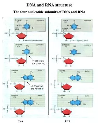
Structure of RNA • Building Blocks • Nucleotide • Ribose sugar • Phosphate group • Nitrogenous bases • Backbone • Alternating Ribose and Phosphate • Nitrogenous Bases • A and G – purines (double ring) • C and U – pyrimidines (single ring) • Usually single stranded • 3 forms • Messenger • Transfer • Ribsomal
Central Dogma • It was determined that RNA serves as an intermediate between genes and proteins. This forms the central dogma of biology - cells are governed by a cellular chain of command: DNA to RNA to protein.
Transcription http://youtu.be/ztPkv7wc3yU • During transcription one of the two DNA strands called the template strand provides a template for ordering the sequence of nucleotides in an RNA transcript. There are three basic stages of transcription: initiation, elongation and termination. These three steps result in the production of messenger RNA (mRNA).
Transcription Initiation begins when transcription factors mediate the binding of the enzyme RNA polymerase to a promoter region onto DNA. The combination of promoter, transcription factors and RNA polymerase is called the transcription initiation complex. A promoter called a TATA box is crucial in forming the initiation complex in eukaryotes. Elongation takes place as RNA polymerase moves along the DNA, unwinding it as it goes (10 to 20 bases at a time.) RNA nucleotides are added (5' to 3' direction) that compliment the DNA template following Charagaff's rule - with one exception -adenine's compliment becomes uracil in the RNA transcript. Termination occurs when the RNA polymerase reaches a special sequence of nucleotides that serve as a termination point. In eukaryotes, the termination region often contains the sequences AAAAAAAA.
Fig. 17-7 Promoter Transcription unit 5 3 3 5 DNA Start point RNA polymerase Initiation Nontemplate strand of DNA 1 Elongation RNA nucleotides 5 3 RNA polymerase 5 3 Template strand of DNA RNA transcript Unwound DNA 3 Elongation 2 3 end Rewound DNA 5 3 5 3 5 3 5 5 Direction of transcription (“downstream”) RNA transcript Template strand of DNA Termination 3 Newly made RNA 3 5 5 3 5 3 Completed RNA transcript
A eukaryotic promoter includes a TATA box 1 Promoter Template 5 3 3 5 TATA box Template DNA strand Start point Several transcription factors must bind to the DNA before RNA polymerase II can do so. 2 Transcription factors 5 3 3 5 Additional transcription factors bind to the DNA along with RNA polymerase II, forming the transcription initiation complex. 3 RNA polymerase II Transcription factors 3 5 5 5 3 RNA transcript Transcription initiation complex
mRNA Editing The 5' end of mRNA receives a "cap" which is a modified gaunine nucleotide that has two additional phosphate groups - forming GTP. Capping provides stability to the mRNA and a point of attachment for the small subunit of the ribosome during translation. The 3' end of mRNA receives a "poly-A tail" which consists of about 200 adenine nucleotides. It provides stability to the mRNA and also appears to control the movement of the mRNA across the nuclear envelope. RNA splicing removes nucleotide segments from mRNA. A transcribed DNA segment contains two kinds of DNA sequence - exons, which are sequences that express a code for a polypeptide, and introns, intervening sequences that are noncoding. Splicing removes introns and joins exons, creating an mRNA molecule with a continuous coding sequence. In some cases, RNA splicing is carried out by spliceosomes. Spliceosomes consist of a variety of proteins and several small nuclear ribonucleoproteins (snRNPs) that recognize the splice sites.
mRNA Editing Alternative splicing allows different mRNA's to be generated from the same RNA transcript. By selective removing different parts of an RNA transcript, different mRNA's can be produced, each coding for a different protein product. Thus the number of different proteins an organism can produce is much greater than its number of genes. Check out the mRNA processing activity in your online textbook. It will help you understand this process.
mRNA • Messenger RNA (mRNA) - is a single strand of RNA that provide the template used for sequencing amino acids into a polypeptide. • A triplet group of three adjacent nucleotides on the mRNA, called a codon, codes for one specific amino acid. • There are 64 possible ways that four nucleotides can be arranged in triplet combinations, there are 64 possible codons. • However, there are only 20 amino acids, and thus, some codons code for the same amino acid. The genetic code, to the right, provides the decoding for each codon. • Of the 64 codons, 61 code for amino acids; 3 triplets are "stop" signals to end translation. • Using the genetic code chart you can determine the amino acid for each codon. For example, if the codon is AUG, the amino acid will be Methionine.
tRNA • Transfer RNA (tRNA) - is a short RNA molecule (about 80 nucleotides) that is used for transporting amino acids to their proper place on the mRNA template. • Molecules of tRNA are not identical, each carries a specific amino acid on one end and has an anticodon on the other end. • The anticodon base-pairs with the complementary codon on mRNA. • Flexible pairing at the third base of a codon is called wobble and allows some tRNAs to bind to more than one codon. • The tRNA molecule looks like a cloverleaf. Because of hydrogen bonds, tRNA actually twists and folds into a three-dimensional molecule. • The correct match between a tRNA and its amino acid is done by the enzyme aminoacyle-tRNAsynthetase. The amino acid attaches to the 3' end of the tRNA molecule.
rRNA • Ribosomal RNA (rRNA) - molecules are the building blocks of ribosomes. The two ribosomal subunits (large and small) are made of proteins and rRNA. • Remember the nucleolus is the area of the nucleus that actively produces/transcribes rRNA and assembles the rRNA together with proteins to form large and small subunits of ribosomes. • Together the two subunits form a complete ribosome. Ribosomescoordinate the activities of mRNA and tRNA during translation. • Ribosomes have four binding sites – • one for mRNA, • one for a tRNA that carries a growing polypeptide chain (P site, for "polypeptide”) • one for a second tRNA that delivers the next amino acid that will be added to the polypeptide chain ( A site, for "amino acid”) • one for a third tRNA that has recently lost its amino acid to growing polypeptide chain and is about to be discharged from the ribosome (E site, for "exit").
Translation http://youtu.be/D5vH4Q_tAkY • During translation mRNA synthesizes polypeptides. This process takes place in three stages: initiation, elongation, and termination. • Initiation - The small ribosomal subunit binds to the 5' end of mRNA and a special initiator tRNA which carries the amino acid methionine. The small subunit moves along the mRNA until it reaches the start codon (AUG). Proteins called initiation factors bring in the large ribosomal subunit that completes the translation initiation complex. The initiator tRNA is sitting in the P site.
Translation • Elongation - During this stage amino acids are added one by one to the preceding amino acid. Each addition involves proteins called elongation factors and occurs in three steps: codon recognition, peptide bond formation and translocation. A new tRNA that complements the mRNA enters into the A site (codon recognition). A peptide bond attaches the amino acid(s) from the tRNA in the P site to the amino acid in the A site (peptide bond formation). The tRNA in the P site no longer has an amino acid attached to it. The ribosome moves down the mRNA strand (translocation) placing the 1st tRNA that has no amino acid in the E site, the 2nd tRNA with the growing polypeptide in the P site, and leaving the A site empty. A new tRNA can now enter the A site. The tRNA in the E site leaves the ribosome. This continues until termination.
Translation • Termination - This process occurs when a stop codon in the mRNA reaches the A site of the ribosome. The A site accepts a protein called a release factor. The release factor causes the addition of a water molecule instead of an amino acid. This reaction releases the polypeptide, and the translation assembly then comes apart.
Phenotypes determined by protein activities… • In 1909, British physician Archibald Garrod first suggested that genes dictate phenotypes through enzymes that catalyze specific chemical reactions • He thought symptoms of an inherited disease reflect an inability to synthesize a certain enzyme • Linking genes to enzymes required understanding that cells synthesize and degrade molecules in a series of steps, a metabolic pathway
One-Gene-One-Enzyme • George Beadle and Edward Tatum exposed bread mold to X-rays, creating mutants that were unable to survive on minimal medium as a result of inability to synthesize certain molecules • Using crosses, they identified three classes of arginine-deficient mutants, each lacking a different enzyme necessary for synthesizing arginine • They developed a one gene–one enzyme hypothesis, which states that each gene dictates production of a specific enzyme • One Gene - One Enzyme
Fig. 17-2 EXPERIMENT No growth: Mutant cells cannot grow and divide Growth: Wild-type cells growing and dividing Minimal medium RESULTS Classes of Neurospora crassa Wild type Class I mutants Class III mutants Class II mutants Minimal medium (MM) (control) MM + ornithine Condition MM + citrulline MM + arginine (control) CONCLUSION Class II mutants (mutation in gene B) Class I mutants (mutation in gene A) Class III mutants (mutation in gene C) Wild type Precursor Precursor Precursor Precursor Gene A Enzyme A Enzyme A Enzyme A Enzyme A Ornithine Ornithine Ornithine Ornithine Gene B Enzyme B Enzyme B Enzyme B Enzyme B Citrulline Citrulline Citrulline Citrulline Gene C Enzyme C Enzyme C Enzyme C Enzyme C Arginine Arginine Arginine Arginine
Mutations • Mutations are changes in genetic material of a cell. Mutations can occur spontaneously or they can be caused by mutagens. • Mutagens are physical or chemical agents that cause mutations. Point mutations are chemical changes in just one base pair of a gene. • Point mutations can be divided into two general categories: • Substitution • Frameshift
Substitutions • Substitution- occurs when the DNA sequence contains an incorrect nucleotide in place of the correct nucleotide. • Silent mutations have no effect on the amino acid produced by a codon because of redundancy in the genetic code. The new codon that results from the mutation codes for the same amino acid. This occurs most often when the nucleotide substitution results in a change of the last of the three nucleotides in a codon. • Missense mutations still code for an amino acid, but not the right amino acid. The effect can be minor, or it may result in a defective protein. The hemoglobin protein that causes sickle-cell disease is caused by a missense mutation. • Nonsense mutations change an amino acid codon into a stop codon, nearly always leading to a nonfunctional protein.
Substitution Mutations Silent Missense Nonsense
Frameshift • Frameshift- occurs as the result of a nucleotide deletion or insertions. Such mutations cause all subsequent nucleotides to be displaced one position. If a frameshift mutations occurs in a DNA segment whose transcription produces mRNA, all codons following the transcribed mutation will change. • deletion - occurs when a nucleotide is omitted to the nucleotide sequence. • insertion - occurs when a nucleotide is added to the nucleotide sequence.
Mutations • Alterations in DNA sequence can lead to changes in the type or amount of protein produced and the consequent phenotype • DNA mutations can be positive, negative or neutral based on the effect or the lack of the effect they have on the resulting nucleic acid or protein and the phenotypes that are conferred by the protein • Mutations are the primary source of genetic variation – especially in prokaryotes