Acute Sinusitis
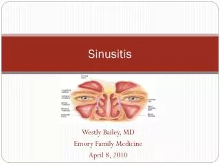
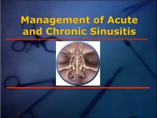
Acute Sinusitis. Anatomy of PNS. A quick recap. There are four paired paranasal sinuses, the maxillary, ethmoid , frontal and sphenoid sinuses “Anterior” and “posterior” sinuses

Acute Sinusitis
E N D
Presentation Transcript
Anatomy of PNS A quick recap
There are four paired paranasal sinuses, the maxillary, ethmoid, frontal and sphenoid sinuses • “Anterior” and “posterior” sinuses • Lining of the sinuses is pseudostratified, columnar epithelium (respiratory epithelium) which is continuous with the nasal epithelium.
The mucosa secretes a mucous which traps bacteria • The mucous is naturally extruded through sinus ostia to be expectorated or swallowed • The drainage of the maxillary and frontal sinuses follows a circular pattern through the natural ostia
Ethmoid Sinuses • Are present at birth, adult size by age 12 • Are separated by the ground (basal) lamella into the anterior and posterior ethmoids, which drain into the middle and superior meatus , respectively
Consist of vertical and horizontal plates • The vertical plate is divided into two portions, the perpendicular plate of the ethmoids and the crista galli • The horizontal plate is known laterally as the fovea ethmoidalis and medially as the cribriform plate • Medially is the lamina papyracea
Maxillary Sinus • The largest sinus • Pyramidal shaped with apex near zygomatic arch • In child, inferior border near nasal floor. In adult, 1 cm below nasal floor • Floor over maxillary dentition, which is often thin and dehiscent over tooth roots
The infraorbital nerve runs along roof, and is often dehiscent. At risk during antral procedures • Sinus ostia located anteriorly in the middle meatus • Accessory ostia are usually more posterior
Frontal Sinus • Rarely present at birth; usually not visible until age 2 • Great variability in size; congenitally absent in 5% • Drains into the frontal recess in the middle meatus near the upper portion of the infundibulum
Sphenoid Sinus • Rarely present at birth, usually seen around age 4 • Drain into the superior meatus in the sphenoethmoidal recess • Ostia of variable size
The optic nerve lies superiorly • The pons lies posteriorly • The cavernous sinus is lateral, along with CNIII, IV and VI and the carotid artery • The carotid artery is dehiscent in 50% of specimens
Lined by respiratory epithelium • Mucous blanket is in two layers: a superficial viscous layer and an underlying serous layer. • Cilia beat in the serous layer, moving the blanket towards the natural ostia • Normal function depends on patent ostia,ciliary function and quality of mucous
Most important pathologic process in disease is obstruction of natural ostia • Obstruction leads to hypooxygenation • Hypooxygenation leads to ciliary dysfunction and poor mucous quality • Ciliary dysfunction leads to retention of secretions
Local factors can impair ciliary function. • Cold air “stuns” the epithelium, resulting in retained secretions. • Dry air dessicates the blanket. • Anatomical factors , polyps, tumors,foreign bodies and rhinitis , block the ostia • Kartagener’s Syndrome (immotile cilia syndrome)
It is usually due to a pyogenic secondary invasion of the mucous membrane after the normal defenses of the nose, the muco-ciliary blanket and lysozyme are breached by an acute virus invasion. • Acute sinusitis following coryza usually affects initially all the sinuses.
It may also occur after trauma or dental extraction. Following trauma such as fractures of the maxilla and frontal bones acute sinusitis may be limited to one sinus and may or may not be associated with a surrounding osteomyelitis. • The commonly found bacteria in this disease include streptococci, staphylococci and pneumococci. Occasionally in post-traumatic cases anaerobic streptococci are found
Inflammation of the mucous membrane of the sinuses leads to oedema and swelling, together with accumulation of exudates and pus cells within the cavity of sinus concerned. • This exudate is ejected through the natural ostium of the sinus by ciliary action. The mucosal swelling and pouring out of fibrin and exudates may block the ostium producing the empyema.
If the body’s defence is poor the infection may spread in to bone; bacteraemia may devlop in to septicemia or pyemia. • Local thrombophlebitis may lead to meningitis, encephalitis, brain abscess or cavernous sinus thrombosis. • The rich blood supply of the area and proximity to the brain and meninges makes the development of intracranial complications a danger.
There is usually a history of coryza followed, after three four days by increasing nasal obstruction and discharge, a deterioration of sense of smell, a sense of discomfort in the face accentuated by bending down and a morning headache.
The ethmoids are the most commonly affected sinuses in acute sinusitis. • The characteristic headache or sensation lies between and behind the eyes.
The swelling of the mucosa may produce a complete loss of sense of smell. • The nasal cavities are intensly congested in the region of middle turbinate and mucopus is seen in the area of ethmoid bulla.
This is less common than maxillary or ethmoid sinusitis. • It is usually unilateral and not infrequently follows swimming or diving when patient has a cold.
Headache over one or the other eye after getting up in the morning, gradually increasing and tending to disappear in the late afternoon. • There is tenderness on pressure over the floor of the sinus. • Little or no swelling of the nasal mucosa may be noticeable, but a trickle of pus may be seen high up at the anterior end of the middle meatus
It usually arises in association with posterior ethmoiditis. • It gives rise to pain which may be severe and is located in the centre of the head. It may some times be referred to the area in front of or slightly above either ear or behind the eyes.
On ant. rhinoscopy purulent discharge may be seen high up in the back of nose between septum & turbinates. • In post-nasal space, pus is seen at the apex of the arch of the posterior choana.
Maxillary sinusitis is characterized by pain in the face on bending down. • There may also be some swelling of the face, frontal headache or pain in the alveolar region. • On examination mucosa of the middle meatus is red and swollen. After sucking out the discharge which is usually present, tilting the head towards the healthy side may produce further discharge.
Occasionally acute maxillary sinusitis follows dental sepsis or extraction. • On examination besides history of dental sepsis or dental extraction, there is marked tenderness and swelling over lower part of the cheek. • Patient may also complain of foul smelling discharge from the nose and foul taste in the mouth. • Pus is seen in the middle meatus and almost every where in the nose.
History & physical signs • Acute sinusitis presents as pain over infected areas, with or without headache • Pain to palpation is common with anterior sinusitis, but is usually absent with the posterior sinuses • Posterior sinuses present as bitemporal or vertex headaches • Fever, malaise, nasal discharge present
Treatment • Acute sinusitis can be thought of as an abscess or empyema • Cornerstone is drainage and antibiotics • Drainage is usually medical with topical decongestants and sometimes antihistamines • In rare cases where medical treatment fails, surgical drainage may be required
S. pneumo , H. flu and M. carrarhalis • Amoxicillin is the first line antibiotic. • Failure to respond to amoxicillin necessitates broading coverage with clavulonic acid and possible Gram’s stain and culture • Surgical drainage is required for failures on augmentin and topical decongestants
Classification • Acute sinusitis is defined as disease lasting less than one month • Subacute sinusitis is defined as disease lasting 1 to 3 months • Chronic sinusitis is defined as disease lasting more than three months, and is usually due to inadequately treated acute or subacute disease
Chronic sinusitis is a chronic inflammation of the mucous membrane which has resulted in irreversible and usually degenerative changes. • The infection may spread to the bony walls.
The organisms usually found are the staphylococcus albus, staph aureus, the Haempphilusinfluenzae and diptheroids. • During periods of more active infection pneumococci and haemolytic streptococci may also be present
Predisposing factors • Anatomical: Irregularities of the nasal septum. • Inadequate pneumatization of sinuses: Attacks of inflammation follow each other in quick succession. The process of inflammation and repair go hand in hand and the resulting scar tissue narrows ostium of the sinus and lead to insufficient aeration. • Hypersensitivity:Allergic changes may be induced by recurrent bouts of acute infection with resulting bacterial hypersensitivity. • Dental sepsis • Inadequate diet • Alcohol and tobacco
Three main categories-1 • Sinusitis associated with simple inflammatory hyperplasia: • It begins in early childhood. Recurrent bouts of infection, shorter periods of remission result in thickened mucous membranes. There is sub-epithelial fibrosis, reduction in glandular tissue. • At a later stage the periosteum becomes affected and hyperemia extends to bone, leading at first to osteoporosis and sclerosis.
Three main categories-2 • Sinusitis as a part of generalized respiratory allergy: • Two types of allergy; generalized allergic diathesis presenting in early childhood. • In second type there is no sign or symptom of allergy till eight or nine years of age after which water logging of mucosa leads to increasing nasal stuffiness and discharge. • Localized edematous areas, perhaps under the pull of gravity, may develop in to polypi especially in ethmoid region.
Three main categories-3 • Either of the two preceding types with superimposed infection. • Allergic subjects are more prone to secondary bacterial invasion than the normal subjects. • Some of the inflammatory products may then act as allergens, reducing further allergic changes.
Clinical Features • There is usually a copious postnasal discharge which may be greenish-yellow. • Nasal obstruction is usually the result of swelling of the inferior turbinate mucosa consequent on presence of sepsis. • Deep chronic headache over the forehead, anosmia or cacosmia. • Secondary symptoms may produce of oedema oeutachean tube orifice, OM, granular phryngitis and chronic laryngitis.
Chronic sinusitis usually seen with a mucopurlentdischarge, but fever is usually not present • Acute sinusitis is often imposed on chronic disease
Diagnosis is primarily clinical, but radiographs can be used • Transilluminationof the sinuses can sometimes be used, but due to differences in sinus size and patency , these tests are not reliable • Antral lavage can be performed in select cases where the diagnosis is in doubt
Treatment • Search for underlying causes which obstruct sinus drainage & ventillation. • C/S of sinus discharge • Initial treatment conservative. • Surgical • FESS