Enzyme Substrate Product
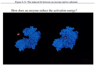
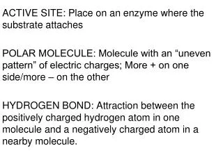
Enzymes. In the microbiology lab, biochemical test relays on enzymes which is glycoprotein or protein that act as catalyst by lowering the activation energy of certain biological reaction.


Enzyme Substrate Product
E N D
Presentation Transcript
Enzymes • In the microbiology lab, biochemical test relays on enzymes which is glycoprotein or protein that act as catalyst by lowering the activation energy of certain biological reaction. • We can use our knowledge in bacterial enzymes to identify the bacteria and distinguish between bacterial species. Enzyme Substrate Product
Types of Enzymes • According to site of the reaction • Endoenzymes : where substrate and enzyme react inside the cell.(ex: oxidase, catalase, urease, nitrate reductase). • Exoenzymes: where substrate and enzyme react outside the cell. (ex: free coagulase, gelatenase, amylase, lipase, casienase). • According to enzyme production • inducible : produced only when needed or induced. • constitutive : produced continuously
Notes: • Every genus of bacteria has it’s unique set of enzymes, so we can identify it. • Endoenzymes may act outside the cell in case of the presence of high concentration of it’s substrate. • The same enzyme could be inducible and constitutive in different genera
Kinds of bacterial enzymatic reactions • The breakdown of toxic wastes such as hydrogen peroxide or urea (ex: catalase) • The reduction of nitrate or oxygen (ex: nitrate reductase). • The degradation of specific amino acids (ex: treptophanase). • The utilization of noncarbohydrate carbon sources for growth (ex: urease).
Catalase Test • Enzyme name \ catalase • Substrate name \ hydrogen peroxide • Enzyme action \ breakdown the toxic H2O2 producing oxygen gas and water • Hydrogen peroxide produce due to the aerobic respiration of the cells and have to be breakdown to prevent it’s toxic action on DNA and cell membrane Catalase 2H2O2 2H2O + O2
When hydrogen peroxide is added to a colony of • catalase-producing bacteria, it is broken down and • the oxygen that is produced can be seen as bubbles. • By catalase test we can distinguish between: • G (+ve) cocci : staphylococcus is catalase positive where streptococcus is catalase negative • G (+ve) bacilli : Bacillus is catalase positive where Clostridium is catalase negative • All Enterobactreacae (a gram negative bacilli) are catalse positive • Lesteria monocytogenes ( a gram positive bacilli) are catalase positive
How to do the Test • Slide method • Add one drop of 3% Hydrogen peroxide on a clean glass slide. • Aseptically take a loopful of the test organism and emulsify in the H2O2 drop. • Capillary tube method • Inoculate the test organism on agar slant and incubate for 24 hours. • Allow 1 mL of 3% hydrogen peroxide to flow over the slant. • Adding hydrogen peroxide directly to a pure slant culture.
Notes: • Be careful when using bacteria from blood agar culture and avoid touching the agar by the loop because blood cell in agar also had catalase enzyme. false positive • Also don’t use bacteria from old culture because the enzymes activity drops by time. false negative
Coagulase Test • Coagulase test is one of the biochemical tests. It is very • important test in the microbiology. The coagulase test • identifies whether an organism produces the exoenzyme • coagulase, which causes the fibrin of blood plasma to clot. • Organisms that produce Coagulase can form protective • barriers of fibrin around themselves, making themselves • highly resistant to phagocytosis, other immune responses, • and some other antimicrobial agents.
Significance • The coagulase test is used to differentiate the potentially pathogenic species Staphylococcus aureus from other Gram-positive cocci, the usually non-pathogenic species. • The S. aureus (potentially pathogenic in humans and animals, but S. epidermidis (is not pathogenic)
Types Of Coagulase • Coagulase enzymes occur in two forms—bound coagulase and free coagulase. • Bound coagulase, also called clumping factor, is attached to the bacterial cell wall and reacts directly with fibrinogen in plasma. The fibrinogen then precipitates causing the cells to clump together in a visible mass. • Free coagulase is an extracellular enzyme (released from the cell) that reacts with a plasma component called coagulase-reacting factor(CRF). The resulting reaction is similar to the conversion of prothrombin and fibrinogen in the normal clotting mechanism.
Test methods • There are 2 methods: • Tube Method (detects the presence of either bound or free). • Slide Method (detects only bound coagulase).
Procedure of Slide Method • Place a drop of coagulase plasma on a clean, dry glass slide. • Place a drop of distilled water or saline near the drop of plasma as a control. • With a sterile loop or wooden stick, emulsify an amount of the isolated colony being tested into each drop. • Inoculating the water or saline first. • Try to create a smooth suspension.
Observe for clumping in the coagulase plasma and a homogenous suspension in the control. Clumps that will not mix uniformly into coagulase plasma indicate a positive test whereas a uniform suspension is indicative of a negative test. • Clumping in both tests indicate that the organism autoagglutinates and is unsuitable for the slide coagulase test. • When autoagglutination is observed.the tube coagulase test should be employed as an alternative to the slide agglutination test.
Procedure of Tube Method • Using a culture that is less than 24 hours old, inoculate the CoaguStaph™ by emulsifying one loopful (2-4 colonies) of bacteria from a non-inhibitory agar plate into the tube of plasma. • Incubate the inoculated tube at 35-37 degrees C. for 1 to 4 hours. • Negative tests at 4 hours should be held at room temperature for a total of 24 hours before reporting results. • Read by gently tilting the tube while observing for clotting of plasma.
Results can be reported across a range 0 to 4+, 0 meaning the plasma remained liquid (no coagulase activity) and 4+ meaning the plasma completely hardened (the consistency of an agar) due to strong coagulase activity. Results should be read at 4 hours. A positive test for coagulase production results in a clotting of the rabbit plasma. Any degree of clotting is a positive test. • All "0" results after 4 hours should be held at room temperature for a total of 24 hours incubation
Notes • When the slide test is employed, all negative slide reactions must be confirmed by the tube test . • The slide agglutination technique may lead to false-positives: • since some strains produce clumping factor resulting in a positive slide test and a negative tube coagulase test. • spontaneous agglutination may occur when rough cultures are used. • The tube test is more reliable than the slide test.
The slide test should be read very quickly, as false • positives can occur. • The slide test should not performed with organisms • taken from high-salt media such as Mannitol Salt • Agar, as the salt content can create false positives.


![Lecture 3: Factors affecting enzyme activity: [substrate] and inhibitors](https://cdn3.slideserve.com/5730998/lecture-3-factors-affecting-enzyme-activity-substrate-and-inhibitors-dt.jpg)