
Digestive System
Digestive System. http://www.umm.edu/imagepages/8710.htm. IB DP Biology Core + Further human Physiology – Option H2 & H3. Digestion Core Material. The Process of Digestion. Large food molecules need to be digested before the nutrients can be absorbed. Why is digestion necessary?.

Digestive System
E N D
Presentation Transcript
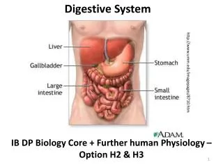
Digestive System http://www.umm.edu/imagepages/8710.htm IB DP Biology Core + Further human Physiology – Option H2 & H3
Large food molecules need to be digested before the nutrients can be absorbed. Why is digestion necessary? Macromolecules are too large to be absorbed though the plasma membrane Polymers are insoluble. Diffusion across membranes requires that Molecules are dissolved in a solution Macromolecules in the food source may not be as useful in the human body., but the component molecules can be digested, absorbed, assimilated, and reassembled into more useful configuration in the body’s cells.
Remember: Hydrolysis (water splitting): breaking down large organic molecules carbohydrate, lipids, and proteins. Enzymes are needed in this process. http://www.doyouknow.in/Articles/Engineering/Types-Of-Chemical-Reactions.aspx
Click to start animation http://highered.mcgraw-hill.com/sites/0072495855/student_view0/chapter2/animation__how_enzymes_work.html
Enzymes are essential in digestion • Enzymes are biological catalysts – globular proteins that increase the rate of a reactions by lowering the activation energy. • Digestive enzymes are released into the gut from glands and are used in catabolic reactions – they break down large molecules. http://upload.wikimedia.org/wikipedia/en/a/af/Catalyst_effect.png • By lowering the activation energy of the reaction, the reaction does not require high temperatures. This is important to living things since high temps would kill them. • By using an enzyme reactions can occur more quickly at body temperature.
Click to start animation Images from: http://programs.northlandcollege.edu/biology/BIOLOGY1111/animations/enzyme.swf
What happens in digestion? Esophagus: wave of muscle contractions (peristalsis) pushes the bolus into the stomach. Mouth: chewing (mechanical digestion) saliva moisten s food to make a bolus of swallowing. Salivary amylase begins chemical digestion of starch. Stomach: muscular contractions continue mechanical digestion. Acid kills bacteria. Pepsin begins digestions of proteins. Duodenum (small intestine): Bile from the liver and gall bladder neutralizes acid and emulsifies fats. Pancreatic amylase and lipase digests carbohydrates and fats. Trypsin digests polypeptides into amino acids. Ileum (mall intestine): Lower half of small intestine absorbs nutrients into the blood via the villi. Large intestine: Water is reclaimed and returned to the blood leaving semi-solid feces. This is stored in the rectum. Egestion: Feces containing undigested food, dead cells, and other waste) is forced out of the anus.
Click to begin animation http://highered.mcgraw-hill.com/sites/0072495855/student_view0/chapter26/animation__organs_of_digestion.html
The stomach Hydrochloric acid in the stomach lowers the pH to around 2, killing bacteria and denaturing proteins. Pepsin enzyme starts protein digestion. Muscular actions aids chemical digestion stretch receptors in the muscular wall trigger release of enzymes. http://www.youtube.com/watch?v=Uzl6M1YlU3w Muscular walls contract for mechanical digestion and mixing enzymes with the food Gastric pits release gastric acid, protective mucus and enzymes. Muscular sphincters control entry of food, exit of chyme (partially digested mixture) Lumen – space in which food is stored while inside the stomach. http://www.umm.edu/imagepages/19223.htm
Small intestine - The small intestine completes digestion of food molecules. • Chyme: entering the duodenum (first section). • Bile:from the liver and gall bladder empties into the duodenum neutralizing the acid and emulsifying fats. • Pancreatic enzymes (amylase, trypsin): are released enzymes are further released into the jejunum http://www.youtube.com/watch?v=bNMsNHqxszc http://microbewiki.kenyon.edu/index.php/Small_Intestine
Small intestine A wave of muscle contractions (peristalsis) keeps the mixture of digested and undigested food moving through the intestine. • The ileum: • Last stage of the small intestine. • Absorption of digested food molecules takes place. • Villi (finger like projections) increase the surface area for absorption and have a rich blood supply. http://microbewiki.kenyon.edu/index.php/Small_Intestine
Absorption and assimilation • Digestion -breaks down large food molecules into smaller ones. • Absorption - uptake of these molecules into the blood& carried to tissues. • Assimilated – taken in to be used. http://www.youtube.com/watch?v=P1sDOJM65Bc Absorption into blood (or lacteals) Assimilated – uptake & used by cells Transport by blood vessels
Label these villus structures and state their function B Villus A http://medical-dictionary.thefreedictionary.com/arachnoid+villi Capillary – Carries blood to and from the villus, maintains concentration gradient. Lacteal – Transports lipoproteins (fats) to circulatory system. Mitochondria – Generates ATP for active transport of food molecules Microvillus – Increase surface area for absorption of food molecules
The Villi are finger like projections in the small intestine which absorb the products of digestion. • Large number of the villi create a huge surface area for absorption of digested food. • Epithelial cells have microvilli – tiny finger like hairs – to further increase surface area. • The single layer thickness of epithelial cells make for a short, efficient diffusion path • A rich blood supply maintains a concentration gradient which nutrients can diffuse down. • Lymph vessels (lacteals) close to the surfaced allow lipids to be easily absorbed. http://missinglink.ucsf.edu/lm/IDS_106_LowerGI/Lower%20GI/mainpages/smallintestine.htm http://missinglink.ucsf.edu/lm/IDS_101_histo_resource/cell_structure.htm
The large intestine • After absorption in the Ileum undigested food is pushed into the Colon (large intestine). • Colon reclaims as much water as possible to the blood before allowing egestion of the solid feces. • Colon - maximizes surface area for absorption of water by being long and folded. • Mucus - secreted to lubricate the passage of feces & muscle contractions keep the feces moving. • Fiber rich diet - clears out waste products & dead cells, reducing the risk of colon cancer. http://ibs.about.com/od/symptomsofib1/ss/Picture-Your-Digestive-System_5.htm
Food passes through the alimentary canal – mouth, esophagus, stomach, small intestine, large intestine, rectum, anus • Gastric juices (fluids containing enzymes) are released into the alimentary canal by various glands: • Salivary • Stomach wall • Pancreas • Small intestine wall http://www.mayoclinic.com/health/medical/IM00374
Do you remember how enzymes work?? http://www.youtube.com/watch?v=E-_r3omrnxw
Exocrine glands: release their secretions into ducts. • Remember: Endocrine glands release their products directly into the blood stream • Digestive exocrine glands: Salivary glands, pyloric glands (stomach), exocrine pancreas, goblet cells (small intestine) • Other examples: Sweat glands, Moll’s gland (eyes), mammary glands, sebaceous glands (skin) Exocrine gland structure can vary from simple tubes to complex arrangements secretory cells acinus lumen Basement membrane http://upload.wikimedia.org/wikipedia/commons/9/99/Gray1105.png
Structures of an exocrine gland cell Rat pancreas - TEM Can you explain why the exocrine gland contain so much RER and Golgi apparatus? http://i-biology.net/
Tight junctions: barriers against the movement of fluid. Tight junctions form between adjacent cells of secretary and absorptive systems (e.g. villi of the small intestine). Adjacent cells are locked together using protein complexes – more protein complexes stronger bond. Junctions prevent movement between cells. All molecules passing to or from the lumen must pass through these cells via diffusion or active transport. This controls what substances pass through. Tight junctions also prevent the movement of molecules from one neighboring cell to another http://en.wikipedia.org/wiki/Tight_junction
Contents of saliva: (about 0.75l produced per day!!) Amylase– start digestion Lingual lipase – break triglycerides into fatty acids Water and electrolytes – moistens & lubricates food Antibacterial compounds…& bacteria too!! http://www.mayoclinic.com/health/medical/IM02997
Stomach lining Contents of gastric juice (pH 1 – 3) • Hydrochloric acid – begins digestion and activates pepsin • Mucus – protects the stomach lining • Enzymes – pepsin and rennin • Gastric juice - produced by the parietal cells in the stomach wall • Production - triggered by detection of peptides. http://www.eytonsearth.org/clay-sleep-disorder.html Parietal cells http://en.wikipedia.org/wiki/Parietal_cell
Contents of pancreatic juice: • Basic (high pH) – bicarbonate ions neutralize acidic gastric juices • Pancreatic juice contains many different enzymes: • Pancreatic lipase • Pancreatic amylase • Trypsin& chymotrypsin (endopeptidases) • Carboxypeptidase & elastase (exopeptidase) http://employee.lsc.edu/faculty/BrianBich/Picture%20Library/Forms/AllItems.aspx?RootFolder=/faculty/BrianBich/Picture Library/Anat-Phys II (Biol 1141)/Endocrine Tissues/Pituitary Gland - Tutorial
Control of gastric secretion: nerves and hormones involved • Gastric secretion is a three phase process: • Cephalic phase: reflex response to visual, smell, or thought stimulus • Gastric phase: four steps • Mechano (stretch) and chemo (protein) receptors send signal • Medulla oblongata receives signal • Exocrine glands produce gastrin in response to signal • More HCl released, pH drops : end result • Intestinal phase: reducing secretions • Low pH or arrival of lipids in the small intestines detected by the medulla oblongata • Gastric secretions inhibited.
The small intestine - digestion and absorption of macromolecules: Ileum Absorption of digested molecules: Surface area maximized by villi and microvilli Duodenum Digestion: close association with pancreas, many enzymes released into the lumen. Membrane-bound enzymes (e.g. maltase) increase efficiency of digestion of some molecules. http://medsci.indiana.edu/a215/virtualscope/docs/chap9_4.htm http://faculty.une.edu/com/abell/histo/histolab3d.htm
Membrane-bound enzymes such as maltase • Some digestive enzymes are locked in place on the epithelium of the villi in the duodenum. • Enzymes are therefore not lost through the digestive tract. • products are released close to the membrane for rapid transport to the blood • digestion of the disaccharides can take place early in the digestive system. • Membrane-bound enzymes that become detached will continue to work in the lumen of the gut. http://i-biology.net/
Cellulose digestion Cellulose is a structural polysaccharide found in foods of plant origin. Digestion of cellulose is by cellulase, which is not produced by humans. Therefore, cellulose passes through the digestive tract and is egested in feces. Cellulose (dietary fiber) remains a vital part of the human diet, as it is aids the health of the digestive system, sweeping out dead cells, unabsorbed materials and bacteria. Wood cellulose http://www.scienceclarified.com/Ca-Ch/Cellulose.html
Digestion of proteins: inactive precursors Pepsin and trypsin are proteases. If they were secreted as active enzymes, they would cause damage to the exocrine cells (auto-digestion). They are instead secreted as inactive precursors (pepsinogen & trypsinogen), which are harmless. The enzymes become activated under the right conditions. http://i-biology.net/
Digestion of proteins: exo- and endo- peptidases http://i-biology.net/ Endopeptidases – (e.g. pepsin, trypsin) hydrolyze bonds in polypeptide chains. They break large polypeptides into smaller ones. Increase surface area for action of exopeptidases. Exopeptidases – (e.g. dipeptidase) remove terminal acids. These amino acids are them available for absorption.
Digestion of lipids: the problem of hydrophobia. Problem: lipids are hydrophobic and form large droplets. Lipases are water soluble so cannot gain access to any more than the outmost lipid molecules in the droplet. Solution: use bile salts to emulsify the lipids. These are secreted by the liver and stored in the gall bladder. Now the fat droplets are broken into smaller droplets with increased surface area. http://i-biology.net/
Stomach ulcers: a major shift in medical treatment The causes of stomach ulcers is an example of how a scientific discovery caused medical experts to change their minds…and lead to a noble prize. For many years it was assumed that stomach ulcers were caused by stress and poor diet. But in the 1980’s it was discovered that Helicobacter pylori (a bacterium) was responsible for most cases of ulcers. H. Pylori infection of the epithelial cells leads to an inflammation and irritation of the lining of the stomach. As a result of this discovery medical practice is now to treat ulcers with a simple course of antibiotics.
Ileum: Absorption of digested molecules Surface area maximized by villi and microvilli http://medsci.indiana.edu/a215/virtualscope/docs/chap9_4.htm
Key absorption points to remember: Microvilli maximize surface area for absorption Tight junctions prevent movement of molecules between neighboring cells. Provide added control of molecule movement. Mitochondria provide ATP for active transport of digested food molecules (glucose, amino acids, mineral ions http://mycozynook.com/102RGCh20OH.htm
Absorption of compounds through the epithelium • Lipids, monosaccharides, & amino acids can diffuse easily across the membrane • Once in the mucosal cell, monoglycerides are converted into lipo-proteins (chylomicrons) in the endoplasmic reticulum. • Exocytosis then delivers them to the lymphatic vessels in the villus, and on the rest of the body. http://bioserv.fiu.edu/~walterm/Fund_Sp2004/digestion/present.htm
Digestion and absorption animations http://nutrition.jbpub.com/resources/animations.cfm# http://nutrition.jbpub.com/resources/animations.cfm#
Fructose absorption through the epithelium: Facilitated diffusion • Fructose and other water-soluble molecules (such as vitamins) move down the concentration gradient into the epithelial cells. • Integral protein channels allow them to pass through the hydrophobic phospholipid bilayer. http://nutrition.jbpub.com/resources/animations.cfm#
Active transport – involved in the movement of amino acids, saccharides, and minerals: • Saccharides e.g.: Glucose, galactose • Minerals e.g.: Calcium, Iron
Some proteins, such as antibodies, need to be absorbed intact. Endocytosis can carry intact molecules. Example: absorption of antibodies from breast milk 3 different types of endocytosis http://apbio12007.blogspot.com/2007_11_01_archive.html Antibodies from breast milk would be brought into the cell by phagocytosis
How does absorption of digested food make use of different methods of membrane transport? http://i-biology.net/


