Download
1 / 26
260 likes | 287 Vues
Explore step-by-step instructions on dissecting a sheep heart, its anatomy, and educational benefits. Ensure classroom safety and maximize students' learning experience.

E N D
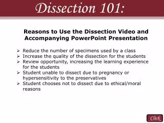
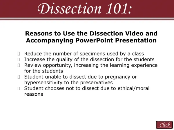
Dissection 101: • Reasons to Use the Dissection Video and • Accompanying PowerPoint Presentation • Reduce the number of specimens used by a class • Increase the quality of the dissection for the students • Review opportunity, increasing the learning experience for the students • Student unable to dissect due to pregnancy or hypersensitivity to the preservatives • Student chooses not to dissect due to ethical/moral reasons Click
Dissection 101: As an educator you are responsible for the implementation of the dissection activity described in the video and PowerPoint. You must have safety procedures and rules established for your classroom and make sure all of the students follow the rules to ensure a safe environment. South Dakota Public Broadcasting and Dakota State University cannot in any way be responsible or liable for any injury as a result of performing the described dissection. Complete the dissection if you feel it is appropriate and safe for your individual class. Have fun and stay safe! • Safety Considerations • Age appropriate activity for the children in your care • Material Safety Data Sheet (MSDS) available for accident reference • Poison control number/phone readily available • Latex free gloves, eye protection and apron/lab coat • Eyewash station, shower and sink • Sharp instruments (cut away from self and others) • Sharps and specimen(s) disposal • Encourage students to report all accidents • Basic science laboratory rules (strictly enforced) Click
Dissection 101: Sheep Heart Click
Sheep Heart Dissection 101: Getting Started • Use water to rinse the excess preservative from your heart Click
Sheep Heart Dissection 101: Sheep Heart The sheep heart is an excellent specimen to use for comparative human anatomy in both size and function. The sheep heart is mammalian, having four chambers like the human heart, which includes two atria and two ventricles. The blood flow through the sheep heart is like that of the human heart, in which the blood is pumped from the right side of the heart to the lungs and then from the left side of the heart to the body. Click
Dissection 101: Sheep Heart Orientation: 1st Example Anterior view (front) Posterior view (back) It is easiest to distinguish the posterior and anterior of the heart by observing the ventricles. Each heart will look a little different, but there are specific things to look for. The following slides show three different hearts. (Also, anatomic left and right are from the viewpoint of the specimen. For example, the left ventricle is an anatomical term describing the ventricle on the left side, but it will appear, many times, on the right side when viewed in books, on your tray, and in this PowerPoint.) Things to look for: Right Ventricle Right Ventricle Left Ventricle Apex Anterior view points right; posterior view points left Posterior interventricular sulcus runs more vertical Anterior interventricular sulcus runs diagonal to the left The left ventricle has more resistance when pressed, because the wall (muscle) is much thicker. Click
Dissection 101: Sheep Heart Orientation: 2nd Example (different heart) Anterior view (front) Posterior view (back) Things to look for: Right Ventricle Right Ventricle Left Ventricles Apex Anterior view points right; posterior view points left Posterior interventricular sulcus runs more vertical Anterior interventricular sulcus runs diagonal to the left The left ventricle has more resistance when pressed, because the wall (muscle) is much thicker. Click
Dissection 101: Sheep Heart Orientation: 3rd Example (different view) The connective tissue and fat have been removed from the heart on the left. Right Ventricle Left Ventricles Right Ventricle Posterior view (back) Anterior view (front) Click
Dissection 101: Sheep Heart Left atrium Structures Right ventricle – Muscular structure of the heart that pumps oxygen poor blood to the lungs Right atrium (atria: plural) – Muscular structure of the heart that pumps oxygen poor blood to the right ventricle Left atrium (atria: plural) – Muscular structure of the heart that pumps oxygen rich blood to the left ventricle Left ventricle – Muscular structure of the heart that pumps oxygen rich blood to the body Auricle – Outer ear like region of the left and right atria Apex – Inferior pointed region of the heart (left ventricle) Anterior view (front) Anterior view (front) tilted down Click
Sheep Heart Dissection 101: There are many vessels entering and leaving the heart. In general, larger vessels leaving the heart are called arteries and larger vessels carrying blood to the heart are called veins. It can be very difficult to identify the vessels, especially if some have been removed by the supplier. On the following slide we will identify vessels which can be easily identified. Many of the other vessels will be identified later with the use of probes that will follow the flow of blood from the heart. Click
Sheep Heart Dissection 101: Brachiocephalic artery – Forms right subclavian artery and common carotid artery; oxygen rich Easily Identified Vessels Aorta – Oxygen rich blood from left ventricle to the body Ligamentum arteriosum – Remnant of ductus arteriosus during fetal development R. A. L. A. Pulmonary Trunk (artery) – Oxygen poor blood from right ventricle to lungs via right and left pulmonary arteries R. V. L. V. Close-up ligamentum arteriosum Click
Sheep Heart Dissection 101: Getting Started – Initial Cuts (YouTube Version) Click
Dissection 101: Sheep Heart Dissection Use probes to identify the superior and inferior vena cava, which supply oxygen poor blood to the right atrium from the body. Right atrium Click
Dissection 101: Sheep Heart Dissection Use the superior vena cava probe, as a guide, to cut down through the wall of the right atrium and right ventricle Right atrium Right ventricle Click
Sheep Heart Dissection 101: Right atrioventricular (AV) valve (also called tricuspid valve) – Prevents backflow of blood into right atrium when right ventricle pumps blood to lungs Right atrium Right ventricle Click
Sheep Heart Dissection 101: Tricuspid valve (right AV valve) Chordae tendineae - Chord like structures (tendon) that connect the tricuspid valve to the papillary muscle Right atrium Papillary muscle – Contracts preventing tricuspid valve from entering the atrium when ventricle pumps blood Right ventricle Click
Sheep Heart Dissection 101: Right ventricle Use a probe to locate the pulmonary trunk Click
Sheep Heart Dissection 101: Once the pulmonary trunk is located, cut the right ventricle along the probe to expose the semilunar valves and pulmonary trunk Click
Sheep Heart Dissection 101: Pulmonary trunk Pulmonary semilunar valve – Prevents backflow of blood from pulmonary trunk into right ventricle Cut right ventricle (probe removed) Close-up view Click
Sheep Heart Dissection 101: Left atrium Left atrium Use a probe to locate the insertion of the pulmonary veins, which bring oxygen rich blood from the lungs to the left atrium; then cut the left atrium along the probe Click
Sheep Heart Dissection 101: Left atrium L. V. Cut along the probe to expose the interior of the left ventricle Probe is passing through the mitral valve into the left ventricle Click
Sheep Heart Dissection 101: Left atrioventricular (AV) valve (also called bicuspid valve or mitral valve) – Prevents backflow of blood into left atrium when left ventricle pumps blood to body Wall left ventricle Left atrium Chordae tendineae Papillary muscles Close-up view mitral valve Click
Dissection 101: Sheep Heart Exposing the Aortic Semilunar Valve (YouTube Version) Click
Sheep Heart Dissection 101: Aorta L. V. Use a probe to locate the aorta; then cut the left ventricle along the probe exposing the aortic semilunar valve Click
Sheep Heart Dissection 101: Aorta Aortic semilunar valve – prevents backflow of blood from aorta into left ventricle Mitral valve Click
Dissection 101: Sheep Heart Produced by Dakota State University and South Dakota Public Broadcasting Science Steve Sponsors