Download
1 / 82
820 likes | 837 Vues
Action and support: the muscles and skeleton. Chapter 40, pages 774-791. Muscles and Skeleton Work Together. Animals—jellyfish, earthworms, crabs, horses, and people—move using the same fundamental mechanism:

E N D
Action and support:the muscles and skeleton Chapter 40, pages 774-791
Muscles and Skeleton Work Together • Animals—jellyfish, earthworms, crabs, horses, and people—move using the same fundamental mechanism: • Contracting muscles exert forces on the skeleton and cause the body to change shape • A body with muscles but no skeleton would not have coordinated movement • A skeleton without muscles remains in one position
Types of Skeletons • Types of skeletons • Hydrostatic skeletons • Exoskeletons • Endoskeletons • Coordinated movement is produced by alternating contractions of antagonistic muscles • Antagonistic muscles act on each type of skeleton to provide movement
Hydrostatic Skeleton • Worms, cnidarians and many mollusks (snails, octopuses) • A hydrostatic skeleton is a sac or tube filled with a liquid • “Hydrostatic” means “to stand with water,” which is how hydrostatic skeletons function • A water-filled balloon “stands up” because it contains water, but if punctured it collapses • The volume of the balloon is fixed, but you can change its shape by squeezing
Earthworm Movement • An animal with a hydrostatic skeleton controls the overall shape of its body using two sets of antagonistic muscles -circular and longitudinal • For an earthworm to move forward it uses wavelike, alternating contractions of longitudinal and circular muscles • Setae hold the front of the worm in place • The worm first contracts its longitudinal muscles in its front end, so it becomes shorter and fatter • Other longitudinal muscles in the middle and tail contract, making it shorter Earthworm Locomotion
The worm contracts circular muscles in its front half, making that half longer and thinner • When the worm is fully extended, longitudinal muscles in the head contract again, fattening and anchoring the head • As the wave of circular muscle contraction moves down the worm, the tail gets thin • Longitudinal muscle contraction in the back half of the worm pulls the tail up toward the head • This cycle is repeated over and over as the worm crawls through the soil
Hydrostatic Skeleton Circular muscles contract Circular muscles relax Longitudinal muscles contract Longitudinal muscles relax liquid liquid (a) Hydrostatic skeleton
Exoskeleton • Arthropods (spiders, crustaceans) and insects, have rigid exoskeletons – outside skeletons • Movement occurs at jointsof the legs, mouthparts, antennae, base of the wings, and body segments • Thin, flexible tissue joins stiff sections of exoskeleton • Antagonistic muscles attach to opposite sides of the inside of a joint, contraction causes movement • Contraction of a flexor muscle bends a joint; contraction of an extensor muscle straightens a joint • Alternating contraction of antagonistic muscles moves the joints
Exoskeleton Extensor muscle contracts Flexor muscle contracts Extensor muscle relaxes Flexor muscle relaxes (b) Exoskeleton
Molting • The exoskeleton cannot expand, an arthropod molt its exoskeleton so that it can grow (27 times in up to 3 yrs)
Endoskeleton • Rigid structures found inside the bodies of echinoderms and chordates • Movement also occurs primarily at joints, where two parts of the skeleton are attached to one another • Biceps - a flexor and triceps – extensor attach on opposite sides of the outside of a joint and move the joint back and forth, or rotate them in one direction or the other
Endoskeleton Flexor muscle (biceps) contracts Flexor muscle relaxes elbow Extensor muscle contracts Extensor muscle (triceps) relaxes (c) Endoskeleton
Functions of Vertebrate Skeleton • Provides a rigid framework that supports the body and protects its internal organs • Allows locomotion • Participates in sensory function • Bones produce red blood cells, white blood cells, and platelets in red bone marrow • Store calcium and phosphorus
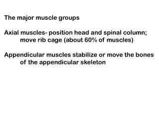
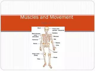
Skeletal Categories • The axial skeleton, which includes the bones of head, vertebral column, and rib cage • The appendicular skeleton – pectoral, pelvic girdles and the appendages attached to them
Structure of Vertebrate Skeletons • Three types of connective tissue—cartilage, bone, and ligament—make up the skeleton • All are living cells embedded in a matrix of collage protein, with various other substances included in the matrix • Bone - larger amounts of minerals composed mostly of calcium and phosphate, and is hard and rigid • Cartilage contains large amounts of glycoproteins and includes elastic fibers, which make some cartilages flexible • Ligaments hold bones together at joints and have small amounts of elastic fibers
Cartilage plays many roles • Provides flexible support and connections • In some fishes (sharks and rays) the entire skeleton is composed of cartilage • During embryonic development, the skeleton (except for skull and collarbone) is first formed as cartilage, and later replaced by bone
More Cartilage Functions • Covers the ends of bones at joints • Supports the flexible portions of the nose and external ear • Provides the framework for the larynx, trachea, and bronchi of the respiratory system • Forms the tough, shock-absorbing intervertebral discs between the vertebrae of the backbone
Cartilage Structure • Chondrocytes are the living cells • Secrete the glycoproteins and collagen that make up the matrix • No blood vessels penetrate cartilage • To exchange wastes and nutrients, chondrocytes rely on diffusion of materials through the collage matrix • Cartilage cells have low metabolic rate, damaged cartilage repairs itself slowly, if at all
Bone • Hard outer shell of compact bone encloses spongy bone • Compact bone is dense and strong, provides an muscle attachment site • Develops as small tubes called osteons with collagen and calcium phosphate surrounding a central canal containing blood vessels • Spongy bone is an open network of bony fibers • Porous, lightweight, rich in blood vessels • Red bone marrow is found in the cavities of spongy bone
Cartilage and Bone cartilage blood vessels central canal spongy bone (contains marrow) chondrocytes osteon compact bone osteocytes collagen matrix
Bone Cells • There are three types of bone cells: • Osteoblasts—bone-forming cells • Osteocytes—mature bone cells • Osteoclasts—bone-dissolving cells • Early in development, when bone replaces cartilage in the skeleton, osteoclasts invade and dissolve the cartilage • Ostoblasts secrete a hardened matrix of bone and gradually become entrapped within it
As bones mature, the trapped osteoblasts mature into osteocytes • Not capable of enlarging a bone • Essential to bone health because they rework the calcium phosphate deposits, preventing excessive crystallization that would make the bone brittle
Bone Remodeling • Allows skeletal repair and adaptation to stress • Each year, 5% -10% of your bone is dissolved. • Replaced by the coordinated activity of osteoclasts that secrete acid and dissolve small amounts of bone, and osteoblasts that secrete new bone • This process allows the skeleton to alter its shape in response to demands placed on it • Bones that carry heavy loads or are subjected to extra stress become thicker, providing more strength and support
Bone remodeling varies with age • Early in life, the activity of osteoblasts outpaces that of osteoclasts, allowing bones to become larger and thicker as a child grows • In the aging body, however, the balance shifts to favor osteoclasts, and bones become more fragile as a result • Although both sexes lose bone mass with age, this is typically more pronounced in women
Broken Bone Repair • The ultimate bone remodeling occurs after a fracture – healing takes about 6 weeks • Typically, the ends of the broken bone are put back into proper alignment • A clot is formed surrounding the broken ends • Cartilage replaces the clot • Bone replaces the cartilage • Completed when mature bone completely replaces cartilage, etc.
Bone Repair 1 2 3 4 Blood from ruptured blood vessels forms a clot surrounding the ends of the broken bone Healing begins when a callus of cartilage replaces the clot Bone gradually replaces the cartilage in the callus When mature bone completely replaces the callus and the original shape of the bone has been mostly restored, the fracture is healed large blood clot compact bone spongy bone
Muscles produce force by contracting • A muscle can only contract or not contract • Muscle lengthening is passive, occurring when muscles relax and are stretched by other forces such as: • contractions of other muscles • weight of a limb • pressure from food • Coordinated movement is produced by alternating contractions of muscles with opposing actions by antagonistic muscles
Structures of Vertebrate Muscles • The muscles of all animals have striking similarities in both the cellular components that produce contractions and in the structural arrangement of these components • The details of muscle structure and function, however, show a tremendous range of adaptations • For example, clams possess a special type of muscle that holds their shells tightly closed for hours using very little energy • Some flies have flight muscles that can contract 1,000 times per second
Types Vertebrate Muscle • Skeletal, cardiac, and smooth • All work on the same basic principles but differ in function, appearance, and control
Skeletal muscle • Moves the skeleton • Cells are striated • Multinucleate • Voluntary or conscious control • Contractions range from quick twitches to powerful, sustained tension • Many nuclei located just beneath the cell’s plasma membrane; largest fibers have several thousand nuclei
Cardiac muscle • Striated • One nucleus per cell • Branched cells • Located only in the heart • Initiates its own contractions, but is influenced by nervous system and hormones • Biofeedback training allows some people to regulate their heartbeat
Smooth muscle • Not striated • Spindle shaped • One nucleus per cell • Surrounds large blood vessels and most hollow organs, producing slow, sustained contractions • Involuntary Control
Skeletal Muscle Cell Structure • Highly organized, repeating structures • Skeletal muscle consists of a series of nested, repeating parts • Skeletal muscles are encased in connective tissue sheaths and attached to the skeleton by tendons • Within the muscle’s outer sheath, individual muscle cells called muscle fibers are grouped into bundles by further coverings of connective tissue
More Details… • Blood vessels and nerves pass through the muscle in the spaces between the bundles • Each individual muscle fiber has its own thin connective tissue wrapping • These multiple connective tissue coverings, each connected to the others, provide the strength needed to keep the muscle from bursting apart during contraction • Muscle cells are among the largest cells in the human body, ranging from 10 - 100 micrometers in diameter and some run the entire length of a muscle, so they can be over 30 centimeters long
Skeletal Muscle Structure connective tissue tendon (connects to bone) nerves and blood vessels bundle of muscle cells muscle fiber (muscle cell) myofibril skeletal muscle
Individual muscle fibers contain many parallel cylinders called myofibrils • Each myofibril is surrounded by a specialized type of endoplasmic reticulum called a sarcoplasmic reticulum • SR is flattened, membrane-enclosed compartments filled with fluid containing a high concentration of calcium ions
The plasma membrane that surrounds each muscle fiber tunnels deep into the inside of the cell at regular intervals, producing tubes called T tubules • T tubules encircle the myofibrils, running between and closely attached to segments of the SR • Each myofibril has repeating subunits called sarcomeres that are aligned end to end along the length of the myofibril, connected to one another by protein discs or Z lines • Within each sarcomere is a precise arrangement of thin and thick protein filaments • Each thin filament is anchored to a Z line at one end • Suspended between the thin filaments are thick filaments
Thin and thick filaments of myofibrils are composed of actin and myosin, they interact to contract the muscle fiber • A myofibril also contains smaller amounts of other proteins hold the fibril together, attach the thin filaments to the Z lines, and regulate contraction • Dystrophinbinds thin filaments to proteins in the plasma membrane, which is are attached to extracellular proteins that surround the muscle fiber • Dystrophin helps to distribute the forces generated during muscle contraction so the fiber doesn’t tear itself apart
Individual actin proteins are nearly spherical • A thin filament consists of two strands of actin proteins wound about each other • Accessory proteins that regulate contraction called troponin and tropomyosin lie atop the actin • A myosin protein is shaped like a hockey stick—a head attached at an angle to a long shaft • The myosin head is hinged to the shaft and can move back and forth
A thick filament consists of a bundle of myosin proteins with a shaft in the middle of the bundle and the heads protruding out • The heads of the two ends of the thick filament are oriented in opposite directions
A Skeletal Muscle Fiber T tubules sarcoplasmic reticulum muscle fiber myofibril plasma membrane (a) Cross-section of a muscle fiber sarcomere myofibril Z lines thin filament thick filament (b) A sarcomere thin filament myosin myosin heads thick filament troponin accessory proteins tropomyosin actin (c) Thick and thin filaments
Skeletal Muscle Contraction • Contraction happens through interaction between thin and thick filaments • The molecular architecture of thin and thick filaments allows them both to grip and to slide past one another, shortening the sarcomeres and producing muscle contraction by what is called the sliding filament mechanism • Each spherical actin protein has a binding site for the myosin head • In a relaxed muscle cell, however, these binding sites on actin are covered by tropomyosin, which prevents the myosin heads from attaching
When a muscle contracts, tropomyosin moves aside, exposing the binding sites on the active proteins • The myosin head binds to these sites, temporarily linking the thick and thin filaments • The myosin heads flex, pulling on the thin filaments and causing them to slide a tiny distance along the thick filament • The myosin heads on the two ends of each thick filament pull the thin filaments toward the middle of the sarcomere
Because thin filaments are attached to the Z lines at the ends of the sarcomere, this movement shortens the sarcomere • All of the sarcomeres of the entire muscle fiber shorten simultaneously, so the whole muscle fiber contracts a little • The myosin heads release the thin filament, extend, reattach farther along the thin filament, and flex again, shortening the muscle fiber a little more, much like a sailor hauling in a long anchor line a little at a time, hand over hand • The cycle repeats as long as the muscle is contracting
The Sliding Filament Mechanism of Muscle Contraction troponin thin filament 1 actin Tropomyosin covers the binding sites, so the myosin head cannot attach tropomyosin binding sites myosin head 4 Using energy from ATP, the myosin head detaches from the actin, extends, and then attaches to another actin binding site farther along on the thin filament myosin (part of a thick filament) 2 When the binding sites of actin are exposed, the myosin head attaches to a binding site myosin head ATP ADP 3 The myosin head flexes, pulling the thin filament past the thick filament and shortening the sarcomere
Muscle contraction requires ATP • Contracting muscles require a lot of energy • One might think that the energy is used to flex the myosin head and pull the thin filament along • The energy of ATP is used not to flex the myosin head, but to extend it and store the energy in this “stretched” position • When the head binds to actin, the stored energy flexes the myosin head and pulls the thin filament toward the center of the sarcomere