Download

1 / 30
300 likes | 732 Vues
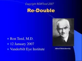
Re-Double Ron Teed, M.D. 12 January 2007 Vanderbilt Eye Institute Alfred Bielschowsky Patient History I cc: vertical binocular diplopia 63 yo male with 4 week history of diplopia; first intermittent, then constant Worse in right gaze No antecedent trauma, CVA, craniofacial surgery

E N D
Re-Double • Ron Teed, M.D. • 12 January 2007 • Vanderbilt Eye Institute Alfred Bielschowsky
Patient History I • cc: vertical binocular diplopia • 63 yo male with 4 week history of diplopia; first intermittent, then constant • Worse in right gaze • No antecedent trauma, CVA, craniofacial surgery • No history strabismus • No history thyroid disease, myasthenia
Patient History II • POH: none • PMH: DJD, hernias • Meds: ibuprofen • FH: no ocular disease • SH: tobacco use in past • ROS: no dizziness, weakness, HA, jaw claudication, fatigue, numbness, paresthesia
Differential Diagnosis of Vertical Binocular Diplopia • Superior Oblique Palsy • Thyroid Ophthalmopathy • Myasthenia Gravis • Brown Syndrome • Orbital fracture with entrapment • Cyclovertical paresis or overaction • Skew Deviation/Ocular Tilt • Dissociated Vertical Deviation
Exam I • General: alert and oriented; no anomalous head posture; no nystagmus • BCVA 20/20, 20/20 • Fields: Full OU • Tonometry: 15,14 • Pupils: no rAPD, no anisocoria • External Exam: no proptosis, ptosis, lid retraction; no fatigue • SLE: unremarkable, quiet eyes • DFE: unremarkable, no optic nerve edema/pallor
Versions 0 0 + 0 0 0 0 0 0 0 0 0
Measurements 0 8 LHT 5 LHT 3 LHT 8 LHT 4 LHT 10 LHT
Additional Clinical Tests • “fourth step” • Measurement of ocular torsion • Double Maddox Rod: 5° excylotorsion OS • Vertical Fusional Amplitudes • Large amplitudes suggest congenital etiology • 3 prism diopters
Superior Oblique Palsy • Clinical diagnosis from Three-step test • What do we do now?
Superior Oblique Palsy • Determine if this is a ISOLATED CN IV palsy • No neurological symptoms on history • Cursory neurological exam unremarkable
Isolated Superior Oblique Palsy • Most common etiologies are congenital and traumatic • Also vascular; less commonly tumor, demyelinating • In absence of other neurological symptoms and presence of vascular risk factors, reasonable to observe
Isolated Superior Oblique Palsy: Management Plan • Our patient did not have obvious vascular risk factors other than age • No known HTN, hyperlipidemia, DM • Patient was observed • To return if diplopia changes, ptosis develops, or he has any numbness, weakness, paresthesias, disorientation, unsteadiness, vertigo, headache
Patient Follow-up • Pt returns 8 weeks later • “double vision is a bit better…” • “…ever since I had the radiation treatment”
Follow Up Exam 0 5 LHT 2 LHT 5 LHT 4 LHT 10 LHT 8 LHT DMR: 5° excylotorsion OS
More History • A few weeks after first visit, pt developed unsteady gait, disequilibrium associated with flank pain • No longer isolated fourth nerve palsy • Measurements no longer map to superior oblique palsy • Now what do we think is going on? • Now what would we do?
Imaging • CT • MRI
Vertical Diplopia and Pontine Mass • Does this lesion explain vertical diplopia? • Lesion to CN IV nucleus or nerve? • Lesion to other pathways encoding vertical gaze?
Back to the original presentation • Was it right to observe an apparent isolated CN IV palsy? • Texts, review articles suggest that observation is acceptable, particularly if the palsy is suspected to be congenital, traumatic, or there is a vascular risk factor • Spontaneous resolution of CN IV palsy occurs within 3 months in 50-95% of patients (better in presumed vascular etiology) • Up to one third have undetermined etiology
Watching the CN IV palsy • “evaluation for an isolated fourth nerve palsy usually yields little information... Older patients should be followed” (BCS, Neuro-ophthalmology) • “MRI…for all patients younger than 45 years with no definite history of significant head trauma, and patients aged 45 to 55 years with no vasculopathic risk factors or trauma” (Wills Eye Manual)
The Evidence • Multiple case series of presumed isolated CN IV palsies • No documented tumors as etiology (Keane 1993: 0/81) • But may fail to adequately confirm true isolation or confirm true CN IV palsy • Lee et al (1998) reviewed cost-effectiveness of imaging • No need to image suspected congenital, traumatic, or vasculopathic palsies
The Rebuttal • A few case reports of isolated CN IV palsies from brainstem strokes • Feinberg and Newman (1999): 6/68 isolated CN IV palsies related to trochlear nerve Schwannoma • Scattered other reports of isolated CN IV palsy from other conditions: • Pituitary macroadenoma • MS, polycythemia rubra
So what do we do? • What is your level of comfort? • How good is your neurological exam? • Reasonable and cost-effective to observe, but you may miss an important lesion
Take Home Points • Determine if an apparent superior oblique palsy is truly isolated • If isolated, it may be reasonable to observe • Understand basic anatomy of the pathways encoding vertical eye movements
References • Brodsky MC, Donahue SP, Vaphiades M, and Brandt T (2006). Skew Deviation Revisited. Survey of Ophthalmology. 51:105-128. • Donahue SP, Lavin PJM, and Hamed LM (1999). Tonic Ocular Tilt Reaction simulating a superior oblique palsy. Archives of Ophthalmology. 117:347-352. • Feinberg AS and Newman NJ (1999) Scwannoma in patients with isolated unilateral trochlear nerve palsy. American Journal of Ophthalmology 127:183-88. • Keane JR (1993). Fourth nerve palsy: Historical review and study of 215 inpatients. Neurology. 43:2439-2443. • Kusher BJ (1989). Errors in the Three-Step Test in the Diagnosis of Vertical Strabismus. Ophthalmology. 96:127-132. • Lee AG, Hayman LA, Beaver HA, et al (1999). A guide to the evaluation of fourth cranial nerve palsies. Strabismus 6(4): 191-200. • Petermann SH and Newman NJ (1999). Pituitary Macroadenoma manifesting as an isolated fourth nerve palsy. American Journal of Ophthalmology 127:235-6. • Thomke F and Ringle K (1999). Isolated superior oblique palsies with brainstem lesions. Neurology. 53(5):1126-27.
CT .
Axial .