DICOM 3D Ultrasound Supplement 43 Overview
320 likes | 2.23k Vues
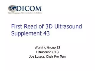
DICOM 3D Ultrasound Supplement 43 Overview Working Group 12 Ultrasound (3D) Joe Luszcz, Chair Pro Tem Goal of Supplement 43 Storage of 3D Ultrasound Volume Datasets, 2D views derived from them, and related information Current Status of Sup 43 Just exited Public Comment phase

DICOM 3D Ultrasound Supplement 43 Overview
E N D
Presentation Transcript
DICOM 3D UltrasoundSupplement 43 Overview Working Group 12 Ultrasound (3D) Joe Luszcz, Chair Pro Tem
Goal of Supplement 43 Storage of 3D Ultrasound Volume Datasets, 2D views derived from them, and related information DICOM 3D Ultrasound Supp 43
Current Status of Sup 43 • Just exited Public Comment phase ftp://medical.nema.org/medical/Dicom/Supps/sup43_pc.pdf • WG-12 (3D) meeting this week to review comments and prepare Letter Ballot draft • Present Letter Ballot draft to WG-6 in March, 2008 DICOM 3D Ultrasound Supp 43
Characteristics of US Modality • Operator-dependent interactive acquisition • Multiple data types possible in each acquisition plane • Non-image data types acquired with image • Lack of a fixed frame of reference • Different probe geometries and oblique acquisition planes • Real-time imaging at relatively high rates DICOM 3D Ultrasound Supp 43
3D Ultrasound Use Cases 1 • Data-related cases • 3D Volume Data • Static and Dynamic Volume Datasets [1,2,22] • Suitable for rendering and slicing [3,4] • Volume calibration [5] • 2D views derived from Volume Data [6,7,8] • Static and Dynamic varieties [9] • Calibrated frame data [10] • Separation of Data Types [11] • Integrate physio waveforms with image [12] DICOM 3D Ultrasound Supp 43
3D Ultrasound Use Cases 2 • Workflow-related cases • Permit Two-Step Review [13] • Frame of Reference • Frame and Transducer-relative FoR • Patient-relative FoR [14,15] • Anatomical FoR (Fiducials) [16] • Key Image identification [20] • 3D Presentation State [17,18,19] • 3D spatial coordinates in SR [21] DICOM 3D Ultrasound Supp 43
Supplement 43 Concepts • Uses same “Enhanced Image” structure used by MR, CT, XA/XRF in which a new Enhanced US Image IOD represents a 3D dataset as a cube of voxels (i.e., a spatial sequence of XY planes) • Data types in separate planes (2D, Flow, etc.) with no embedded graphics or text • Transducer Frame of Reference (primary) with optional Gantry and Patient Frames of Reference • New Enhanced Blending and Display Pipeline for combining data types for display • Uses companion Waveform IOD’s for related physio waveforms • Derived 2D images represented by US Image IOD’s DICOM 3D Ultrasound Supp 43
Ultrasound 3D Image Types DICOM 3D Ultrasound Supp 43
Why a role for 2D US IOD’s? • Ease the transition for 3D ultrasound images on legacy PACS • Provide separate SOP Classes for two-stage query/retrieval • Maintain images with integrated data types for direct presentation on viewers • Defer the work of generalizing Enhanced US for 2D images DICOM 3D Ultrasound Supp 43
Not in Supplement 43 • Line or frustum data representations (if desired, use linked Raw Data IOD) • Use of Enhanced US IOD as 2D replacement (future activity) • Oblique volume datasets (oblique views relegated to US-MF IOD) • Attempts to standardize rendering and slicing algorithms (may differ in appearance) DICOM 3D Ultrasound Supp 43
What is covered by Sup43? • New Enhanced US Image IOD for 3D volume datasets • Single (“3D”) or Multiple temporally-related volumes (aka “4D”, “3D loop”, “3D animation”) • “Slices” consisting of one or more frames of distinct data types at same space and time • 3 new Waveform IOD’s • Frame of reference for hand-held transducer as well as gantry and patient Frames of reference DICOM 3D Ultrasound Supp 43
What is covered (continued) • MONOCHROME2 data representation • New Enhanced Blending and Display Pipeline defines grayscale and/or color presentation for single or multiple data types • Support for multi-palette color LUTs for data frames in the same image • New Ultrasound Application Profiles • Carry-over of ultrasound exam context from existing US image IOD’s, many as Code Sequences DICOM 3D Ultrasound Supp 43
Q & A DICOM 3D Ultrasound Supp 43
Slice Registration DICOM 3D Ultrasound Supp 43
Separation of Data Types DICOM 3D Ultrasound Supp 43
Use of Multi-Frame Dimension • Attributes defining data type alignment use existing Multi-frame Dimensions • Typical order of significance: • Stack ID (most significant) • Temporal Position Index • In-Stack Position • Data Type (least significant) DICOM 3D Ultrasound Supp 43
Common Values Sequence • New “Common Values SQ” provides high-level view of dimensional symmetry • For each dimension, indicates those attributes that share the same value in every frame with the same value of the dimension attribute • Augments, but doesn’t replace “Per-Frame Functional Groups” DICOM 3D Ultrasound Supp 43
Transducer Marker Orientation Attribute specifying the orientation of a “tactile marker” relative to the image data. Enumerated values: • POS_X the image is oriented such that the tactile marker is in the direction of positive X • NEG_X the image is oriented such that the tactile marker is in the direction of negative X DICOM 3D Ultrasound Supp 43
US Frames of Reference DICOM 3D Ultrasound Supp 43
Patient Frame Of Reference DICOM 3D Ultrasound Supp 43
Patient FoR Transformation Transducer Gantry Position and Orientation attributes containing a translation vector SXYZ and 3 “directional cosines” XXYZ, YXYZ, and ZXYZ indicating the position and orientation of the Gantry FoR relative to the Transducer FoR according to the following transformation: DICOM 3D Ultrasound Supp 43
Representation of Color Images • “MONOCHROME2” photometric interpretation used for all Enhanced US Image data values • Presentation State used to associate palette color LUT’s for color representation (colorized tissue or flow/TDI data) • Types combined in the new Enhanced Blending and Display Pipeline DICOM 3D Ultrasound Supp 43
Blending and Display Pipeline DICOM 3D Ultrasound Supp 43
Is Blending for 2D or 3D? Written in terms of 2D Data Frames, but it is applicable to • any 2D result of a slicing or rendering operation that preserves individual data types • transformation into RGB volume prior to slicing or rendering operation DICOM 3D Ultrasound Supp 43
Physio Waveforms Conveyed in separate Waveform objects… • Linked with the image object via bi-directional SOP Instance UID references • Synchronized via Acquisition Datetime values in a common time reference • With each waveform type in a separate Waveform SOP Class • With gated acquisition info via R-Wave identification through the Waveform Annotation Module and excluded beats identified via the Volume Timing module, permitting our “Live 3D stacked ECG display” DICOM 3D Ultrasound Supp 43
Stacked ECG Display DICOM 3D Ultrasound Supp 43
Volume Timing DICOM 3D Ultrasound Supp 43
Known Open Issues 1) Section C.8.X.1: Coverage of 3D Stress and improved 2D Protocols (Staged and Non-Staged) is intended. Does this provide enough flexibility for so-called “custom protocols”? 2) Section C.8.X.1: Not sure what should be included in “Protocol Context” or its Modifier. [added v25] 9) The Enhanced Palette Color Lookup Table describes an improved Enhanced Blending and Display Pipeline. Is the level of complexity correct (flexible enough, yet implementable in readers). Please review this pipeline to verify whether the flexibility is sufficient yet not excessively complex. [added v31] 10) No new Presentation State SOP Class has been defined to reference the Enhanced Blending and Display Pipeline. [added v31] 11) Add authoritative references for sample frequency range for Arterial Pulse Waveform, Respiratory Waveform and Phonocardiographic Waveform. [added v32] 12) What are the appropriate numbers of channels and sample frequencies for the new Pulse, Respiration, and Phonocardiologic Waveform IODs? Are the Waveform Sample Interpretation attribute values correctly defined (linear, logarithmic, etc.)? [added v33] DICOM 3D Ultrasound Supp 43
More Known Open Issues 13) Should the Enhanced US IOD require the use of the VOI LUT module or macro? [added v33] 14) Would a blending pipeline with symmetric primary and secondary path be more helpful in terms of easier implementation considering that it would be implementation of symmetric path or would that increase the complexity of the pipeline without much value to it? [added v33] 15) What should be the media application profile for Ultrasound? Should the General Purpose media profile be used, or should the existing Ultrasound application profile be extended for Enhanced Ultrasound object, as appears in this draft? [added v35] DICOM 3D Ultrasound Supp 43
More Known Open Issues 16) The Enhanced US Image Module (Section C.8.X.3) includes a number of optional attributes from the existing US Image Module (ps3.3 Section C.8.5.6). Is it appropriate to keep the following Type 3 attributes in the Enhanced US Image Module, and if so, is the form correct (defined terms vs. code sequence values, for example)? • Beat Rejection Flag (0018,1080) • Transducer Data (0018,5010) • Focus Depth (0018,5012) • Preprocessing Function (0018,5020) • Mechanical Index (0018,5022) • Bone Thermal Index (0018,5024) • Cranial Thermal Index (0018,5026) • Soft Tissue Thermal Index (0018,5027) • Soft Tissue-focus Thermal Index (0018,5028) • Soft Tissue-surface Thermal Index (0018,5029) • Depth of Scan Field (0018,5050) 17) The Transducer Type attribute (0018,6031) attribute has been replaced with the two code sequence attributes: Transducer Type Code Sequence (0018,xx09) and Transducer Type Modifier Code Sequence (0018,xx0A), with corresponding Context Groups CID xxx05 and CID xxx06. Is this information necessary? Is this scheme acceptable and are the choices in the Context Groups correct and complete? [added v35 and modified v38] DICOM 3D Ultrasound Supp 43