I .Sinus Rhythms and Sinus Arrhythmias
650 likes | 1.34k Vues
I .Sinus Rhythms and Sinus Arrhythmias. 1. ECG of Sinus Rhythms Sinus rhythm must originate in the sino-atrial node. 1) .Regularly recurring sequences of P waves, QRS complexes,and T waves.P-P or R-R interval establishes a specific interval which should not vary more than 0.12 second.

I .Sinus Rhythms and Sinus Arrhythmias
E N D
Presentation Transcript
I.Sinus Rhythms and Sinus Arrhythmias • 1.ECG of Sinus RhythmsSinus rhythm must originate in the sino-atrial node.1).Regularly recurring sequences of P waves, QRS complexes,and T waves.P-P or R-R interval establishes a specific interval which should not vary more than 0.12 second.
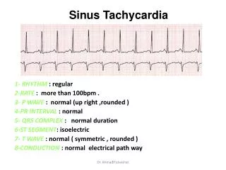
I.Sinus Rhythms and Sinus Arrhythmias • 2).The P wave is upward in lead I,II, avF,V4-5 and downward in lead avR.3).The PR interval>0.12 second.4).Heart rate between 60 and 100 rates per minute.
I.Sinus Rhythms and Sinus Arrhythmias • 2. Sinus Tachycardia1). 1).2).and 3)2).Heart rate exceeding 100 per minute .
Factors associated with Sinus Tachycardia:Physiologic Exercise Strong emotion Pain Anxiety states
I.Sinus Rhythms and Sinus Arrhythmias • PathologicFeverHyperthyroidismHemorrhageShockAnemiaInfectionCongestive heart failureMyocarditisHypoxia
I.Sinus Rhythms and Sinus Arrhythmias • Other factorsDrugs Epinephrine Atropine Food,etcTea coffeeAlcoholTobacco
I.Sinus Rhythms and Sinus Arrhythmias • 3.Sinus Bradycardia1).1),2) and 3).2).Heart rate is less than 60 per minute.
Sinus Rhythms and Sinus • Arrhythmias • Common causes • Physiologic bradycardiaLaborers and trained athletesEmotional states leading to syncopeCarotid sinus pressure, eyeball pressure,intracranial pressureSleep
PathologicSystemic diseaseObstructive jaundiceObstructive diseases of the intestine,kidney or bladderDuring convalescence after some diseases marked by fever(e.g.influenza)myxedemamyocardial infarction(inferior wall or atrial infarction)high intracranial pressure
I.Sinus Rhythms and Sinus Arrhythmias • DrugDigitalisMorphineQuinidinePropranolol
I.Sinus Rhythms and Sinus Arrhythmias • 4.sinus arrhythmia (1) 1) .2) .3)and 4) (2) P-P or R-R interval varies in duration by at least 0.12 second
I.Sinus Rhythms and Sinus Arrhythmias • Common Causes Active rheumatic fever Infectious diseases Atelectasis Chronic adhesive pleuritis Intracranial tension Digitalization Autonomic nerve (It is normal in children and young adults.)
I.Sinus Rhythms and Sinus Arrhythmias • Note: It varies with the phases of respiration,the Sinus rate increasing with inspiration and decreasing with expiration.
I.Sinus Rhythms and Sinus Arrhythmias • 5.Sinus arrest There is no sinus P wave in ECG suddenly.The long interval is not times of P-P interval.
II.Premature beat • The terms “premature beat”,”premature contraction”,”premature systole”,or “extrasystole” indicate that the atria ,AV junction, or ventricle are stimulated prematurely.
II.Premature beat • These premature beats are called “atrial premature beats”when they arise in some portion of the atria .AV junctional premature beats arise in the AV junction. Ventricular premature beats arise in one of the branches of the bundle of His ,the Purkinje network ,or the ventricular muscle.
II.Premature beat • 1. Ventricular premature beats1).The QRS complex is premature ,is 0.12second or more wide ,and is aberrant,notched ,or slurred .It is associated with a T wave that usually point in a direction opposite to the main deflection of the QRS complex.2).The premature QRS complex is not preceded by a P wave.
II.Premature beat • 3).A ventricular premature beat is often followed by a fully compensatory pause(the sum of the R-R intervals including the pre-premature beat and the post-premature beat interval equals the sum of two normal R-R intervals)4).Multiply, ventricular premature beats that arise from a single focus show a similar shape and usually a similar coupling intervals (distance from the preceding normal QRS complex to the premature ventricular beat) in any one lead.
II.Premature beat • 5).occasionally, a ventricular premature beat will occur simultaneously with the apex of the preceding T wave,This is R on T phenomenon. When this occurs ,it may be a precursor of a ventricular tachycardia. • Note: multifocal ventricular prematyre beat (VPB) and multiformed VPB
II.Premature beat • 2.Atrial premature beats1).A premature P wave is present .It may be surperimposed on the preceding T wave because it is premature.The premature P wave is usually followed by a QRS complex and a T wave.Occasionally, it is not followed by a QRS complex and a T wave .(blocked atrial premature beat).2).The QRS and T waves that follow the premature P waves usually resemble the other QRS and T waves in the lead.
II.Premature beat • 3).The P-R interval of the atrial premature beat is usually longer than the normal PR intervals in the ECG.4).An atrial premature beat is often followed by a noncompensatory pause.5).The ventricular complex is usually normal but may be aberrant in from if the premature atrial beat coincides with the refractory phase of the previous ventricular beat .The aberrant QRS is called aberrant conduction.
II.Premature beat • 3. AV Junctional premature beats 1).A premature AV junction P wave is followed by a QRS and T wave.2).The AV junction P waves in aVR become upward .The P waves in II,III, and aVF is downward.The PR interval is usually less than 0.12second ,if the P waves is before the QRS complexes. The P waves may appear after the QRS complexes or may be hidden within the QRS complex.3).An AV junctional premature beat is followed by a fully compensatory.
Ⅲ.Ectopic tachycadia • It is more common to paroxysmal tachycardia. The paroxysmal tachycardia can be divided into two main groups.① Paroxysmal Supraventricular tachycardia② Paroxysmal ventricular tachycardia
Ⅲ.Ectopic tachycadia • 1.paroxymal supraventricular tachycardiaECG 1).Heart rate is regular rhythm with a rate o f 160-250/minute.2).The QRS complex in form is usually normal.3).The P wave in not easy to see.4).With abrupt onset and abrupt terminal.
Ⅲ.Ectopic tachycadia • 2. paroxysmal ventricular tachycardia1).The QRS complex are 0.12 second or more wide ,are aberrant ,and are followed by aberrant ST segments and T waves.2) Ventricular rate is between 140 and 200/minute and regular rhythm or slightly irregular.3).The P waves have no relation to the QRS complexes.4).Fusion beats or ventricular capture are present.5).Sometimes, P-P interval >R-R interval.but the P-R is no relation.
Ⅳ.Flutter and Fibrillation • The flutter and fibrillation arise from excitable ectoptic focus in the atria and ventricle and with a rapid rate and appropriate conduction block. Thus ,They are easily caused by a reentry.
Ⅳ.Flutter and Fibrillation • 1. Atrial FlutterECG: 1).There are no P waves in ECG 2).Presence of saw-tooth flutter wave.3).F waves always uniform in size ,shape and frequency.4).Regular atrial rhythm with a rate of 250-3505).Ventricular response of 1:1,2:1,3:1,4:1,or higher.6).Absence of isoelectric line.
Ⅳ.Flutter and Fibrillation • 2. Atrial FibrillationECG: 1).Absence of P waves2).P waves replaced by f waves.3).f waves : irregular in size ,shape ,and spacing. Rate between 350 and 6004). Irregularly irregular ventricular rhythm, best seen in Ⅱ,Ⅲ,Avf,V1 or V2.
Ⅴ.Atrio –ventricular block(AVB) • AV block, or heart block, exists when conduction of the stimulus from the atria to the ventricle through the AV node is slowed or blocked.The AV block may be transient ,intermittent ,or permanent .It may be incomplete or complete. A patient may show various types of AV block in one ECG.
AVB • 1. First degree heart block(Ⅰ゜AVB)I゜AVB is prolongation of the atrio-ventricular conduction time and is also referred to as first degree A-V block.ECG:prolonged P-R interval:longer than 0.20sec in adults and >0.22s in old adults.The difference of P-R interval between two times is more than 0.04 second.Note:P-R interval varies with heart rate and age.
AVB • 2.II゜AVB (second degree heart block)1).Mobitz Type I(Wenckeback phenomenon)(1)The P-R interval becomes longer and longer (2)The R-R interval gets shorter and shorter, until there is a blocked or nonconducted ventricular beat with a long pause, then an escape rhythm or beat resumes.
AVB • 2).II゜II type(mobity type II AV block) Mobity II is characterized by failure of conduction of one or more sinus beats to the ventricle .There is a fixed numerical relationship between atrial and ventricular impulses,which may be 2:1 or 3:1 or 4:1 .Mobitz II blocks become progressive worse until a complete heart block is established.Thus ,mobitz Type II require a pacemaker,whereas mobitz I does not require a pacemaker,since it does not progress to complete heart block.
AVB • 3.III゜AVB(Complete heart block) (Third degree A-V Block)ECG:1).The atrial and the ventricular rhythms are absolutely independent of one another .2).There is no P-R to QRS relationship.3).The atrial rate is more rapid than the ventricular rate.4).regular P-P interval .5).rugular R-R interval
AVB • 6).QRS is 0.12sec or greater. VR is 36 beats per minute or less.(20-40 beats/mim)QRS is less than 0.12sec.VR is 36 to 60 beats per min(40-60beats/min)