Download
1 / 18
500 likes | 2.92k Vues
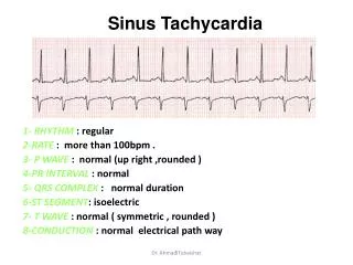
Sinus Tachycardia. 1- RHYTHM : regular 2-RATE : more than 100bpm . 3- P WAVE : normal (up right ,rounded ) 4-PR INTERVAL : normal 5- QRS COMPLEX : normal duration 6-ST SEGMENT : isoelectric 7- T WAVE : normal ( symmetric , rounded )

E N D
Sinus Tachycardia 1- RHYTHM : regular 2-RATE: more than 100bpm . 3- P WAVE : normal (up right ,rounded ) 4-PR INTERVAL : normal 5- QRS COMPLEX : normal duration 6-ST SEGMENT: isoelectric 7- T WAVE : normal ( symmetric , rounded ) 8-CONDUCTION: normal electrical path way Dr. Ahmad Tubaishat
Sinus Bradycardia Dr. Ahmad Tubaishat
1- RHYTHM: rate increased with inspiration ,decreased with expiration 2-RATE: varies between 50 -100 bpm . 3- P WAVE: normal (up right ,rounded ) 4-PR INTERVAL: normal 5- QRS COMPLEX: normal duration 6-ST SEGMENT: isoelectric 7- T WAVE: normal ( symmetric , rounded ) 8-CONDUCTION: normal electrical path way Sinus arrhythmia Dr. Ahmad Tubaishat
Sinus Arrest Dr. Ahmad Tubaishat
Atrial Flutter Dr. Ahmad Tubaishat
Atrial Fibrillation Dr. Ahmad Tubaishat
Paroxysmal Supraventricular Tachycardia (PSVT) • 1- RATE: between (140 – 200) bpm . • 2- RHYTHM : R-R interval regular . • 3- P WAVE : absent or fused with QRS or T wave . • 4- P-R INTERVAL : absent . • 5- QRS COMPLEX : narrow < 0.06 second . • 6- T WAVE : peaked T wave . • 7- CONDUCTION : the ventricles is stimulated from some where in the atria Dr. Ahmad Tubaishat
Premature Ventricular Contraction • Arise within an ectopic focus within the ventricle (no atrial activity). • Vent. Conduction spreads more slowly through purkinje system wide QRS • No preceding P wave, T wave opposite direction of the QRS. • Multiform PVCs: different contours; Multifocal PVCs: different origin • Bigeminy: one normal QRS fowled by PVC; Trigeminy: 2 sinus QRS fowled by PVC; Quadrigeminy: 3 sinus QRS fowled by PVC • Two PVCs in row: couplet, three in row: triplet (a short run of VT) Dr. Ahmad Tubaishat
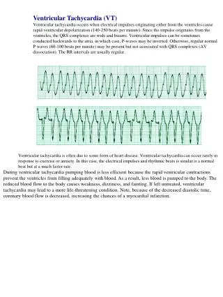
Ventricular Tachycardia Dr. Ahmad Tubaishat
Ventricular Fibrillation Dr. Ahmad Tubaishat
Junctional Rhythm Lead II Dr. Ahmad Tubaishat
Premature Junctional Contraction Ectopic impulse from a focus in the AV junction, occur prematurely, before the next sinus impulses. Dr. Ahmad Tubaishat
First-degree AV block • Prolongation of AV conduction • P wave: present and precedes each QRS • PR: constant but exceeds the upper limit (>0.2second) • Rate: 60 -100 bpm • Rhythm: regular with constant prolonged PR interval Dr. Ahmad Tubaishat
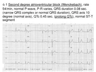
Second-degree AV block, Mobitz type I • One or more of the atrial impulses fail to reach the ventricles • Progressive prolongation of PR interval flowed by missing QRS • Rate: 60 -100 bpm • Rhythm: regular atrial, irregular ventricular Dr. Ahmad Tubaishat
Second-degree AV block, Mobitz type II • One or more of the atrial impulses fail to reach the ventricles • Constant normal PR interval flowed by missing QRS • The block occur occasionally or in 2:1, 3:1 or 4:1 fashion • Rate: 60 -100 bpm • Rhythm: regular atrial, irregular or regular ventricular depends on the AV block Dr. Ahmad Tubaishat
Second-degree AV block, Mobitz type II • One or more of the atrial impulses fail to reach the ventricles • Constant normal PR interval flowed by missing QRS • The block occur occasionally or in 2:1, 3:1 or 4:1 fashion • Rate: 60 -100 bpm • Rhythm: regular atrial, irregular or regular ventricular depends on the AV block Dr. Ahmad Tubaishat
Right Bundle Branch Block Dr. Ahmad Tubaishat
Left Bundle Branch Block Dr. Ahmad Tubaishat