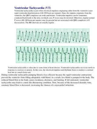
Ventricular Tachycardia
Ventricular Tachycardia. PBL 1. VT - Definition. A general term that applies to a ny rhythm faster than 100 bpm arising distal to the bundle of HIS. Sustained VT with haemodynaic instability (syncope, hypotention ) is life threatening. Based on ECG, can be either: Monomorphic VT

Ventricular Tachycardia
E N D
Presentation Transcript
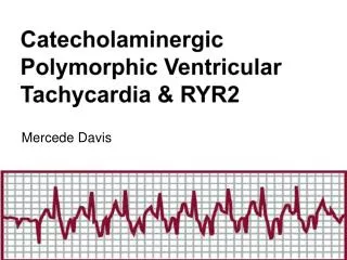
Ventricular Tachycardia PBL 1
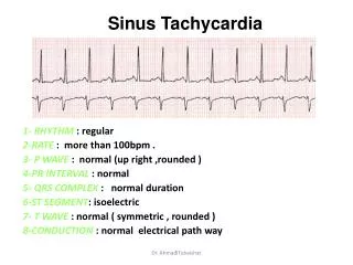
VT - Definition • A general term that applies to any rhythm faster than 100 bpm arising distal to the bundle of HIS. • Sustained VT with haemodynaic instability (syncope, hypotention) is life threatening. • Based on ECG, can be either: • Monomorphic VT • Polymorphic VT • Can also be classified as ‘sustained’ (>30 secs) or ‘non-sustained’
VT polymorphic monomorphic VT Non-sustained sustained • Kumar and Clark • Life threatening ventricular tachyarrhythmias • Sustained ventricular tachycardia • ventricular fibrillation • Torsades de pointes • Normal heart ventricular tachycardia • Non-sustained ventricular tachycardia
Monomorphic VT • Occurs when the ventricular activation sequence is the same, resulting in a uniform ECG pattern.
Monomorphic VT – Structural abnormalities • Most typically seen with patients with a structural heart abnormality. • Usually there is a region of slow conduction • Causes include scarring and fibrillar disarray due to: infract, cardiomyopathy, hypertrophy, surgical scar and muscle degeneration. • Re-entrant tachycardias occur when there is scar tissue or an abnormal area that causes the electrical wavefront to move through it slowly. By the time it reaches the normal (fast) conducting area has already repolarized, causing it to depolarize again and start a loop.
Monomorphic VT – non structural abnormalities • A minority of pt with monomorphic VT have structurally normal hearts (idiopathic VT). • Usually a benign condition with good prognosis. • The VT arises from either a focus in the right ventricular outflow tract or in the left ventricular septum. • In symptomatic pt, radiofrequency catheter ablation of the area causing the VT results in a cure in >90% pt.
Polymorphic VT • The ECG trace shows a varying pattern. • Causes very broadly include: • Drug induced • Inherited and Cardiac ion channel defects • Ischaemia and myocarditis (most common causes when no drug and ion channel defects)
Polymorphic VT - Torsades de pointes • Torsades de pointes is a polymorphic VT following a long resting long QT interval (>0.44 seconds). The ECG waves are rapid, and irregular (i.e. polymorphic) and continuously change from an upright to inverted position.
Torsades de pointes • Causes include (congenital vs acquired) • Congenital – Long QT syndrome: Jervell-Lange-Nielsen or Romano-Ward syndrome. • Acquired: • Electrolyte abnormalities – hypokalaemia, hypomagnesaemia, hypocalcaemia • Drugs – quinidine, erythromyocine, haloperidol, disopyramide, etc. • Misc – bradycardia, mitral valve prolapse, AMI Pathophysiologyis complex – related to ionic flow, which leads to broad QT, which then leads to early after depolarisations.
Torsades de pointes • Clinical presentation: • Causes palpitations, syncope and dizziness, but usually spontaneously resolves. • It can lead to VF and sudden death • Acute management – • Any electrolyte imbalances corrected • Cauasative drugs stopped • Heart rate maintained with atrial or ventricular pacing. • Magnesium sulfate 8mmol over 10-15mins. This stops early after depolarisations through causing an influx of Ca.
Torsades de pointes - Long term management • Acquired – • Avoidance of all drugs known to prolong QT interval • Congenital – • Beta blockade • Left cardiac sympathetic denervation • Pacemaker therapy
Polymorphic VT - others • There is a list of other syndromes caused by different channels being affected, Kumar and Clark only highlights: • Brugada Syndrome – inherited condition that leads to patients having idiopathic ventricular fibrillations with no evidence of causative structural cardiac disease. • Only successful treatment in symptomatic patients is ICD.
Brugada syndrome • Characteristic ECG: right bundle branch block (rsR pattern in V1 with T wave inversion) with ST elevations in V1 –V3
Brugada syndrome • Patient may present with: sudden death during sleep, resuscitated cardiac arrest and syncope or they may be asymptomatic and their ECG may lead to diagnosis. • High risk of sudden death. • Only successful treatment is ICD
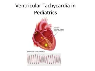
Sustained Ventricular Tachycardia • Presentation • Sustained ventricular tachycardia (>30secs) often with: presyncope, syncope, hypotention and cardiac arrest. Nb: some pt may tolerate it very well. • Ex. • 120-220bpm • Often signs of atriaoventricular dissociation – cannon ‘a’ waves in neck
Ix: ECG • Broad (>0.12) abnormal QRS complexes. May have some capture beats (intermittent narrow QRS complex from normal ventricular contraction via AV node)
NB: 80% of all broad complex tachycardias are due to VT. Other causes include SVT with either a right or left bundle branch block. • Mx: • If patient haemodynamically compromised (i.e. Is hypotensive, has pulmonary oedema) emergency DC conversion may be needed. • If blood pressure and CO are maintained, IV therapy with class 1 drugs used: lidocaine.
VT vs SVT with BBB • VT more likely if • A very broad QRS>0.14s • AV dissociation • A bifid, upright QRS with a taller first peak in V1 • A deep S wave in V6 • Same polarity QRS direction in all chest leads v1 – v6.
Non-sustained VT (NSVT) • > than 5 consecutive beats, but lasts less than 30 secs. • Found in up to 6% of pt with normal heart and does not require treatment. • Found in up to 60-80% of pt with heart disease. In patients with particularly poor left ventricular function (ejection fraction <30%) ICD may help.
VT polymorphic monomorphic VT Non-sustained sustained Kumar and Clark Life threatening ventricular tachyarrhythmias Sustained ventricular tachycardia ventricular fibrillation Torsades de pointes Normal heart ventricular tachycardia Non-sustained ventricular tachycardia