Download

1 / 18
180 likes | 451 Vues
FOOT ISCHEMIA FOLLOWING FOAM SCLEROTHERAPY. G. Lionel Zumbro, Jr., MD, FACS, RVT, RPVI. HISTORY. 57 yo female underwent RF ablation of left GSV with multiple phlebectomy. Ten days later she had RF ablation of the right GSV.

E N D
FOOT ISCHEMIA FOLLOWING FOAM SCLEROTHERAPY G. Lionel Zumbro, Jr., MD, FACS, RVT, RPVI
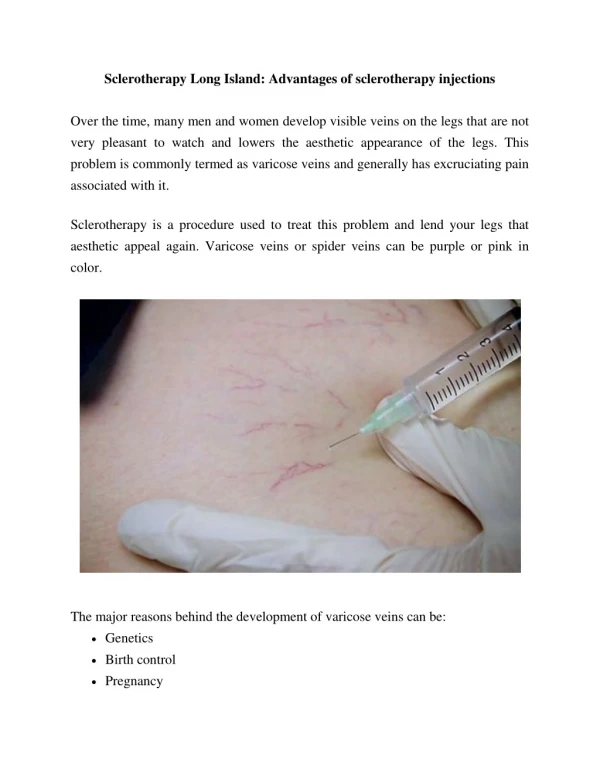
HISTORY • 57 yo female underwent RF ablation of left GSV with multiple phlebectomy. • Ten days later she had RF ablation of the right GSV. • Concurrent sclerotherapy of the right anterior ankle and dorsum of the foot VV. • 0.5% STS CO2/O2 foam, 3 cc. • US confirmed foam in VV. Elevated and wrapped with Coflex. Foot and toes looked good postop.
POD 1 Patient returned C/O thigh tenderness. Foot pulses good. Capillary refill good in toes. • POD 6 Returned with foot and toes dusky. Still good pulses. Digital plethysmography showed no pulse 2nd and 5th toes. Hospitalized. Arteriography.
treatment • IV Heparin, Intraarterial TPA, + Verapamil • ASA, Coumadin, Hyperbaric O2
Patient discharged after 5 days hospitalization. No tissue loss. • Eight months later: “Doctor, you injected the Plantar Arch Artery”.
Shunt flow of arteriovenous fistulas from plantar artery Y Yoshida and M Fujita Human Health Sciences, Kyoto University Graduate School of Medicine, Kyoto, Japan __________________________________________________________________________________________ Abstract Objective: The purpose of this study was to visualize the shunt flow of arteriovenous fistulas (AVFs) passing towards the top side of the foot from the plantar artery. Methods: Colour-flow duplex Doppler ultrasonography was performed in 112 patients who consulted an outpatient clinic with varicose veins and/or symptoms such as foot oedema, dullness, cramp and coldness. Thirteen age- and sex-matched healthy subjects served as controls. Results: AVFs were detected in 86 of 112 patients (77%). They were also detected in 10 (77%) of 13 healthy subjects. The shunt flow pattern consisted of two phases of flow corresponding to systole and diastole, and the diastolic fraction of time– velocity integral was larger, although the peak flow velocity in systole was higher than that in diastole. Conclusion: Colour-flow duplex Doppler ultrasonography is useful for non-invasive visualization of the shunt flow of AVFs connecting the plantar artery with the venous arch of the top side of the foot. Phlebology 2011;26:32-34
Colour Doppler ultrasonographic image of an arteriovenous fistula (AVF). Red signal shows an AVF flow into the venous arch of the top side of the foot from the plantar artery. Yoshida Y, Fujita M. Shunt flow of arteriovenous fistulas from plantar artery. Phlebology 2011;26:32-34
Flow pattern of an arteriovenous fistula. Biphasic flow velocity pattern was obtained with the pulse-wave Doppler ultrasonography. Yoshida Y, Fujita M. Shunt flow of arteriovenous fistulas from plantar artery. Phlebology 2011;26:32-34
Blood flow velocity of the dorsalis pedis artery before and after footbath. Yoshida Y, Fujita M. Shunt flow of arteriovenous fistulas from plantar artery. Phlebology 2011;26:32-34
Perfect storm • Elevation – Empty • Foam to feet • Heat – Wrapping • Pressure – Wrapping • Long term venous hypertension • Venous to arterial shunting
Conclusions • AV shunts do exist • Chronic venous hypertension probably affects shunt flow • Pulses, capillary filling and lack of pain is misleading • Treatment can be effective