Chronic Visceral Ischemia
390 likes | 648 Vues
Chronic Visceral Ischemia. Hallmarks. Abdominal pain after eating (15-20 min) Mikkelsen - Intestinal angina Abdominal pain post food ingestion Increasing frequency and severity Changes in types, amounts and frequency of food eaten 90% pts. Have lost weight (29 lbs.)

Chronic Visceral Ischemia
E N D
Presentation Transcript
Hallmarks • Abdominal pain after eating (15-20 min) • Mikkelsen - Intestinal angina • Abdominal pain post food ingestion • Increasing frequency and severity • Changes in types, amounts and frequency of food eaten • 90% pts. Have lost weight (29 lbs.) • 75% have an abdominal bruit
Historical Perspectives • Described 1918 - Baccelli • Conner 1933 - Misdiagnosed abdominal pain • Dunphy (Peter Bent Brigham Hospital) 1936 - Precursor to fatal intestinal necrosis • Mikkelson 1957 proposed treatment • 1958 Shaw and Maynard - first successful TEA of the SMA
Epidemiology • 4:1 - Female:male • 60.7 y/o +/- 10 years • 16% do not have classic symptoms • nausea, vomiting, diarrhea, constipation • 17.4 mo. +/- 12.2 mo delay in diagnosis • 50% have had some form of abdominal operation
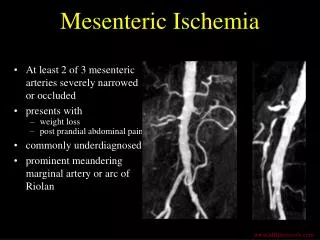
Etiology • Near 100% have occlusion or high grade stenosis of the SMA • 70% have disease of all 2-3 mesenteric vessels • 20-30% have disease of the SMA or celiac alone ???? • Atherosclerosis • FMD, Arteritis, Median arcuate ligament
Incidence • 6-10% Unselected autopsy patients have significant atherosclerosis of visceral arteries • 14-24 % on Aortogram
Diagnosis • Biplanar aortography • MRA • CT scan with IV contrast • Duplex ultrasonography • Other • Upper GI, Endoscopy Plain films • 33% present for other Vascular dz. manifestations
MRA • 2D-TOF MRA coronal plane • Splenic artery, trunks of SMA and Renal artery • NOT GOOD FOR: • Hepatic artery, gastroduodenal artery, SMA branches • Kumamoto Japan, 1995
MRA (plus) • Gadolinium enhanced MRA • 65 patients/14 positive MRA • 12 Angio alone/2 Surgery alone • 6 patients had mesenteric ischemia • Stenosis/occlusion of the Celiac in 7 pts. • Stenosis/occlusion of the SMA in 6 pts. • 100% sensitive/95% specific; 100% sensitive • 1997, University of Michigan • 1998, Universitat Heidelberg
MRA (plus-plus) • Gadolinium enhanced MRA (before and after caloric stimulation) • 10 Healthy volunteers (28 second breath hold) • Fasting (6 hours)/Fed 475 kcal meal • SMA flow 2.3-7.3 ml/min/kg • SMV flow 3.4-9.1.ml/min/kg • Clearly enhances distal vessels better • 1998, University Hospital Zurich
Duplex Scanning • Celiac and SMA have an anterior position • Hampered by: bowel gas,obesity • Used best in: Normal subjects, response to stimuli, Postoperative evaluation • SMA - reversed flow in early diastole (high resistance) in fasting state (Not true in celiac) • SMA PSV-103-196 cm/sec/PDV 15.8-55 cm/sec - higher PDV in the celiac
SMA and Celiac Hemodynamics • 15 min - 90 min post meal • SMA increase in PSV<increase in PDV • Celiac velocity not as fed state dependent • Celiac responds before SMA • Glucagon very similar to post fed state • Vasopressin • Decreased PSV and PDV • Celiac >> SMA
Collateral Circulation • SMA Occlusion • Hepatic to GDA • Superior pancreaticoduodenal to inferior pancreatic
Collateral Circulation • Celiac occlusion • Reverse flow • IMA to SMA • Meandering mesenteric • Mid colic to ascending colic branch • Iliac to IMA • Sup/Inf Hemorrhoidal
Duplex Scanning • 80 patients (Total) • n=9 SMA stenosis > 70% • PSV > 275 cm/sec, no postprandial scanning necessary • 89% sensitivity • PSV<275 cm/sec, postprandial increase > 20% • 100 % specificity that SMA dz. Is not present • 1995, Oregon Health Sciences (Porter)
Intervention Indications • Symptomatic patients with appropriate biplanar angiography • Negative wokup for other sources of pain • Asymptomatic patients at time of aortoiliac reconstruction with SMA stenosis/occlusion • SMA alone - ? Retrograde bypass • SMA and celiac - Supraceliac bypass
Intervention(What to revascularize) • Late recurrence 26.5% (n=56 pts) • 11% with complete revascularization • 29% with 2/3 revascularization • 50% with 1/3 revacularization • Mayo Clinic 1981 (Hollier) • Similar results 1 or 2 arteries, as long as SMA revascularized • UCSF (Reilly), Oregon Health Sciences (Porter)
Interventions • Transaortic endarterectomy • Antegrade bypass • Retrograde bypass • Vein patch angioplasty • Interpostion graft • PTA • PTA/Stent
Visceral Endarterectomy • Blind retrograde fashion • During acute ischemia in sick patient • Distal SMA • Incision through the origin of the SMA
Transaortic endarterectomy • 50% of patients atheroma is around SMA/celiac orifice • 10-20% had to have conversion to another procedure • Medial visceral rotation • Combined aortic, renal and visceral repair • 14.6% peri-op mortality, 17% multiple complications • UCSF, 1991 (Cunningham)
Transaortic endarterectomy • Usually works because lesions represent “overflow disease” • Distal disease (SMA) • Vein patch angioplasty
Antegrade Bypass • Lower morbidity (less extensive surgery) • 95.8% 1 year/ 86% 5 year relief of symptoms • Slightly shorter ischemia times (26 vs. 30 min) • UCSF, 1991 (Cunningham)
Antegrade Bypass • N=16 • 100% some component of organ failure • 25% peri-op mortality of MODS • University of Florida 1987 (Harward)
Antegrade Bypass • Transabdominal • Incise the arcuate ligament • Expose the aorta > 6cm proximal to the celiac axis • Celiac artery proximal to its branches, common hepatic artery, SMA at or beyond the ligament of Trietz • Retropancreatic tunnel
Retrograde Bypass(Infrarenal Aorta) • More disease in the distal aorta • IMA - easy prograde flow • SMA - must put the graft high on the aorta, and distally on the SMA • Celiac - requires a long graft • Very prone to kinking, thrombosis, compression • *Note: Follow up studies have not shown this problem clearly
Retrograde Bypass(Infrarenal Aorta) • Short vein graft vs. long vs. prosthetic • All lengths can kink • Vein 500 ml/min max (SMA = 750 ml/min) • Vein is preferential in cases of bowel necrosis
Retrograde Bypass(Infrarenal Aorta - Right Iliac junction) • SMA alone • Dissect ligament of Treitz • Graft comes through mesentery to anterior SMA wall • SMA and Celiac • Bifurcated graft • Celiac limb - Retropancreatic to hepatic artery
PTA • Poor surgical candidates • 1980 - Furrer • Poor results with “orificial” lesions • Symptomatic relief 80% at 3 year • 5% mortality, 20% failure, 15 % - 50% need recurrent therapy • 40% recurrent sx at 1 year
PTA • Better outcome if oversized balloons used • Symptomatic relief not related to the pressure drop, may be related to overdilation • Southampton Hospital 1988 (Odurny) • Baylor 1996 (Allen)
PTA with Stent • 12 patients/3 celiac artery stenosis, 7 SMA stenosis, 1 aortomesenteric bpg occlusion, 2 SMA occlusions • 92% technical success • 8% mortality despite technically successful procedure • Primary patency 74%, secondary patency 83% at 3 years • Rhode Island 1999, (Sheeran)
Evaluation of Intervention Patency • Intra-op Duplex imaging • Intra-op Electromagnetic flow measurement • Routine post-op angiogram • Some consider madatory
Peri-operative care • Pre-operative TPN • Post-opertive TPN • “Revascularization syndrome” • Abdominal pain, tachycardia, leukocytosis, intestinal edema
Celiac Artery Compression Syndrome • Clinicopathologic condition vs. anatomic condition • 80% female • Disease: Reproducible postprandial pain, age 40-60, > 20 lb. Weight loss • Anatomic: Atypical remitting pain, Psch d/o, substance abuse, >60 y.o., weight loss < 20 lb.
Evaluation • Abdominal pain, abdominal bruit and appropriate arteriogram (> 50% lesion) • Upper and lower GI series • HIDA • ERCP • Small bowel biopsy • Abdominal CT • Malabsorption studies • UCSF 1985 (Reilly)
Interventions • Decompression • 53% remained asymptomatic • Decompression and dilation • 76% remained asymptomatic • Reconstruction • 76% asymptomatic • *Arteriogram: Asymptomatic patient -70% wide open celiac, Symptomatic - 75% celiac stenosis
TAKEHOME • In the right hands most techniques are comparable • TEA - if multiple vessels need revascularization • Antegrade vs. retrograde - depending on pts’ anatomy, concomminant procedures
TAKEHOME • PTA • Fibromuscular disease • Poor operative risk patients • Older patients • Stent ostial lesions • Necrotic bowel • Vein bypass • Direct thromboendartectomy
TAKEHOME • Median Arcuate Ligament Syndrome • Be exhaustive in patient workup • Asymptomatic disease in patients having infrarenal aortic surgery