Download
1 / 69
1.16k likes | 2.81k Vues
Urine concentration and dilution. Learning objectives. Describe the mechanism of formation of dilute and concentrated urine.

E N D
Learning objectives • Describe the mechanism of formation of dilute and concentrated urine. • Describe the role of the ascending limb of the loop of Henle in producing a high renal interstitial fluid osmolality. Beginning with the loop of Henle, contrast the tubular fluid and interstitial fluid osmolality changes that allow either dilute or concentrated urine to be produced and excreted.
Learning objectives • Identify the tubular section and cellular mechanism by which ADH increases permeability to water and urea. • Distinguish between central and nephrogenic diabetes insipidus based on plasma ADH levels and the response to an injection of ADH.
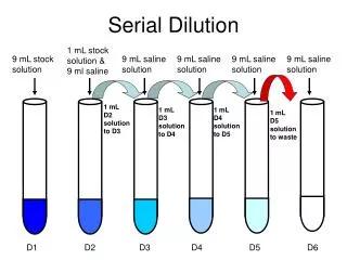
1. Because the osmolarity of the interstitial fluid of the renal medulla becomes progressively greater, more and more water is reabsorbed by osmosis as tubular fluid flows along the descending limb toward the tip of the loop.
2. Cells lining the thick ascending limb of the loop have symporters that actively reabsorb Na, K, and Clfrom the tubular fluid . As solutes—but not water molecules—are leaving the tubular fluid, its osmolarity drops to about 150 mOsm/liter. 3. While the fluid continues flowing along the distal convoluted tubule, additional solutes but only a few water molecules are reabsorbed.
4. Finally, the principal cells of the late distal convoluted tubules and collecting ducts are impermeable to water when the ADH level is very low.
Formation of Concentrated Urine • The ability of ADH to cause excretion of concentrated urine depends on the presence of an osmotic gradient of solutes in the interstitial fluid of the renal medulla
Solute concentration of the interstitial fluid in the kidney increases from about 300 mOsm/liter in the renal cortex to about 1200 mOsm/liter deep in the renal medulla. • The three major solutes that contribute to this high osmolarityare Na, Cl , and urea. • Two main factors contribute to building and maintaining this osmotic gradient: (1) differences in solute and water permeability and reabsorption in different sections of the long loops of Henle and the collecting ducts, and (2) the counter current flow of fluid through tube-shaped structures in the renal medulla.
Overview of Countercurrent Multiplication Mechanism • Descending limb extracts water and concentrates the ultrafiltrate • Thick ascending limb extracts NaCl • Concentrates medullary interstitium • Dilutes ultrafiltrate
Generating a Concentrated Medullary Interstitium - Key Steps Cortical Collecting Duct Urea Cortex 4 3 2 5 H2O 1 H2O Na+ Outer Medulla K+ NaCl 2Cl- H2O Medullary Collecting Duct NaCl H2O Inner Medulla NaCl Urea NaCl Urea H2O NaCl Urea NaCl Loop of Henle
Counter current multiplication • Is the process by which a progressively increasing osmotic gradient is formed in the interstitial fluid of the renal medulla as a result of countercurrent flow.
PT DT 300 300 300 300 300 300 300 300 300 300 300 300 300 300 300 300 300 300 300 300 300 300 300 300 300 300 LH Countercurrent Multiplication - Schematic Model Initial Conditions: Assume all fluid starts out isotonic
200 400 400 200 400 400 200 400 400 200 400 400 200 400 400 400 200 400 200 400 400 200 400 400 Countercurrent Multiplication - Schematic Model PT DT Flow Stopped: NaCl and H2O movements occur (assume 200 mOsm/L gradient is established by thick AL pump) LH
200 300 200 300 200 300 200 300 400 400 400 400 400 400 400 400 Countercurrent Multiplication - Schematic Model PT DT 200 300 Flow Occurs: Fluid leaves ascending limb and new fluid enters descending limb from PT LH
150 350 350 150 350 350 150 350 350 150 350 350 300 500 500 500 300 500 300 500 500 300 500 500 Countercurrent Multiplication - Schematic Model PT DT Flow Stopped: NaCl and H2O movements occur LH
150 300 150 300 300 350 300 350 300 350 350 300 500 500 500 500 Countercurrent Multiplication - Schematic Model PT DT 150 300 Flow Occurs: Fluid leaves ascending limb and new fluid enters descending limb from PT LH
125 325 325 125 325 325 225 425 425 225 425 425 225 425 425 425 225 425 400 600 600 400 600 600 Countercurrent Multiplication - Schematic Model PT DT Flow Stopped: NaCl and H2O movements occur LH
125 300 225 325 225 325 225 425 225 425 425 400 400 425 600 600 Countercurrent Multiplication - Schematic Model PT DT 125 300 Flow Occurs: Fluid leaves ascending limb and new fluid enters descending limb from PT LH
112 312 312 175 375 375 175 375 375 225 425 425 225 425 425 513 313 513 313 513 513 500 700 700 Countercurrent Multiplication - Schematic Model PT DT Flow Stopped: NaCl and H2O movements occur Etc., etc.! LH
Countercurrent Exchange • Is the process by which solutes and water are passively exchanged between the blood of the vasa recta and interstitial fluid of the renal medulla as a result of countercurrentflow
Slow flow: • Flow through the loop is relatively slow. This is also a characteristic of flow through the capillary loops (vasa recta). Increased tubular fluid flow diminishes the ability of the loop to generate and maintain the osmolar interstitial gradient. • Increased flow through the vasa recta washes out the metabolites. • Loss of the high medullaryosmolarity reduces the ability of the kidneys to form a concentrated urine
Q ADH will be released from the posterior pituitary when there is a decrease in a. Plasma Na+ concentration b. Plasma volume c. Plasma K+ concentration d. Plasma pH e. Plasma Ca2+concentration
Water Handling in the Collecting Duct - ADH Absent 18 L No ADH Osm 300 Osm 50 H2O H2O H2O H2O H2O Osm 50 Aquaporin 2 18 L Osm 1200
Osm 300 Osm 1200 Water Handling in the Collecting Duct - ADH Present 18 L ADH Osm 50 H2O H2O H2O H2O H2O H2O Osm 1200 Aquaporin 2 0.5 L
Production of concentrated urine by the kidneys occurs in the following way : 1) Symporters in thick ascending limb cells of the loop of Henlecause a buildup of Naand Cl in the renal medulla. 2) Countercurrent flow through the descending and ascending limbs of the loop of Henle establishes an osmotic gradient in the renal medulla
tubular fluid becomes progressively more concentrated as it flows along the descending limb and progressively more dilute as it moves along the ascending limb. 3) Cells in the collecting ducts reabsorb more water and urea.{ under ADH} 4 )Urea recycling causes a build up of urea in the renal medulla
Q. In the presence of ADH, the filtrate will be isotonic to plasma in the a. Descending limb of the loop of Henle b. Ascending limb of the loop of Henle c. Cortical collecting tubule d. Medullary collecting tubule e. Renal pelvis
Q • What is the term used to describe the amount of pure water that would be added or removed from urine per unit time in order to make it have the same osmolarity as plasma?
Free-water clearance (CH2O) • is used to estimate the ability to concentrate or dilute the urine. • Free water, or solute-free water, is produced in the diluting segments of the kidney (i.e., thick ascending limb and early distal tubule), where NaCl is reabsorbed and free water is left behind in the tubular fluid. • In the absence of ADH, this solute-free water is excreted and CH2O is positive. • In the presence of ADH, this solute-free water is not excreted, but is reabsorbed by the late distal tubule and collecting ducts, and CH2O is negative.
Q What kidney ability is free water clearance actually a measure of? FWC is measure of the ability of the kidney to excrete water
Q. What kind of urine generate a negative value for free water clearance? Hyperosmotic urine Q. What kind of urine generate a positive value for free water clearance? Hypo osmotic urine
Q What kind of urine generate a value of zero for free water clearance? • iso osmotic urine • Positive-free water clearance tends to cause increased plasma osmolarity; • negative free water clearance causes reduced plasma osmolarity.
FWC = 1 – urine osmolarity × V plasma osmolarity Where V is volume of urine per unit time
Q • V = 3.0 mL/min • Uosm = 800 mOsm/L • Posm = 400 mOsm/L What is the free water clearance?