Download
1 / 32
320 likes | 477 Vues
Functional neuroanatomy and plasticity of the hypothalamic circuits regulating autonomic responses to stress. Krisztina J. Kovács Laboratory of Molecular Neuroendocrinology Institute of Experimental Medicine Budapest, Hungary. CHRONIC or UNRESOLVED. HPA Sympato-medullar activity

E N D
Functional neuroanatomy and plasticity of the hypothalamic circuits regulating autonomic responses to stress Krisztina J. Kovács Laboratory of Molecular Neuroendocrinology Institute of Experimental Medicine Budapest, Hungary
CHRONIC or UNRESOLVED HPA Sympato-medullar activity Sympato-adrenal Heart rate Blood pressure Respiratory rate Muscle tension Plasma glucose Gastrointestinal actvity ACUTE STRESS Metabolic X syndrome Mood swings Anxiety Depression Confusion / forgetfulness Burnout Eating disorders Sleeping disorders Social withdrawal / aggression Drug abuse
Selye explains STRESS – induced activation of HPA axis Nature, July 4, 1936. A Syndrome produced by Diverse Nocuous Agents “…. a typical response appears, the symptoms of which are independent of the nature of the damaging agent… and represent rather a response to damage as such” H. Selye
PARAVENTRICULAR NUCLEUS Parvocellular part Magnocellular part Hypophyseotropic Autonom projection • Medial parvocellular dorsal • Periventricular • Medial parvocellular ventral • Dorsal parvocellular • Lateral parvocellular
PVN Brain stem & spinal cord BAT AVP OXY CRH HEART SKINSUDOMOTOR ADRENAL MEDULLA CRH & AVP HPA AXIS ACTIVATION VASCULAR TONE AVP & OXY OSMOREGULATION CARDIOVASCULAR REGULATION
Nociceptive “Blood borne” Somatosensory Visual Visceral Acustic Corticosterone Afferent connections of the hypothalamic PVN
Challenge-induced Activation of PVN Neurons Kovács et al, 2005 c-Fos-ir 90 min after stress*
Dynorphin CRH Angiotensin II CCK Angiotensin II Enkephalin Enkephalin CCK Dopamine TRH Enkephalin Galanin Neurotensin Galanin VIP/PHI VIP/PHI Colocalization of neuropeptides in the hypothalamic PVN PARVOCELLULAR MAGNOCELLULAR VASOPRESSIN OXYTOCIN CRH ? Oxytocin CRH Vasopressin Somatostatin Dynorphin Enkephaline Vasopressin
Functional plasticity in the PVN - adrenalectomy Control Adrenalectomy ADX + DEX/PVN
Vasopressin potentiates CRH action at the corticotropes Rivier et al, 1984
ETHER STRESS-INDUCED VASOPRESSIN TRANSCRIPTION IN THE PARAVENTRICULAR NUCLEUS
Autonomic projection neurons in the PVN Approx. 1500 neurons in 3 different parvocellular subdivisions: (dorsal-, ventral aspect of medial parvocellular- and lateral-) Neurochemical specificity of these neurons is less known: (Oxytocin, vasopressin, corticotropin-releasing hormone, somatostatin, dynorphin, enkephalin, dopamine…..) Efferent connections: to medullar and spinal preganglionic cells for both divisions (sympathetic and parasympathetic) of the ANS- spinal cord (intermediolateral cell column) - predominantly OXY dorsal vagal complex - predominantly AVP Express ER-beta, MC4R, IRS-2 etc Physiological evidences for mediation of sympatoexcitation...
FUNCTIONAL PLASTICITY OF HYPOTHALAMIC AUTONOMIC-RELATED NEURONS
+ sucrose + / - sucrose ADX / SHAM Record body weight, fluid and food consumption Blood sampling perfusion adaptation 1. wk 2. wk 3.wk 4.wk 5. wk 6. wk CRH mRNA in situ AVP mRNA in situ Adult male Wistar rats1M sucrose, 0.5% NaCl, water
Sucrose ingestion results in neuronal activation in neuroendocrine and autonomic-related neurons
Hypothalamus coordinates autonomic responses in part through AVP, released in NTS. AVP inhibits afferent synaptic transmission in the NTS: 1. By decreasing glutamate release probability (V1a receptor) 2. By inducing synaptic failures and increased conduction times
Trans synaptic tracing using pseudorabies virus (PRV) Trans-synaptic spread) PRV: pseudorabies virus, Bartha strain “self-amplifying transsynaptic tracer”.
Virus-infected neurons in the PVN following inoculation into the kidney
Virus-infected neurons in the dorsal parvocellular subdivision
PRV-ir neurons after virus inoculation into a peripheral target 5. Insular cortex 4. Hypothalamus, PVN 3. A5 noradrenergic cell group 2. Rostral ventrolateral medulla • Spinal cord, • intermediolateral cell column
Comparison of autonomic innervation of WAT and BAT Double-virus infection WAT BAT
PRV injections Ba-Dup-Lac (red)- iWAT Ba-Dup-Green- BAT
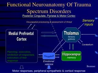
Outline of the brain circuit that provides sympathetic innervation of different target tissues Five cell groups in the brain appear to regulate the entire sympathetic outflow: the paraventricular hypothalamic nucleus (PVH), A5 noradrenergic cell group, caudal raphe region, rostral ventrolateral medulla, and ventromedial medulla. Target organ
PITUITARY AP PP PREGANGLIONIC NEURONS IML DVC CORTEX LIMBIC CORTEX PVN BNST Amygdala Medial parvo Magno Auton. related OXY, VP, CRH DYN, ENK CRH, VP Parabrachial A5 VP, OXY ACTH ADRENAL CORTEX MEDULLA ENDOCRINE AUTONOMIC BEHAVIORAL