Download
1 / 12
120 likes | 308 Vues
Functional Neuroanatomy of Human Rapid-Eye-Movement Sleep and Dreaming. Pierre Maquet , Jean-Marie Peters , Joel Aerts , Guy Delfiore , Christian Degueldre , Andre Luxen , & Georges Franck . Sharon Dhaliwal. Introduction. What is the purpose of this study?

E N D
Functional Neuroanatomy of Human Rapid-Eye-Movement Sleep and Dreaming Pierre Maquet, Jean-Marie Peters , Joel Aerts , Guy Delfiore, Christian Degueldre, Andre Luxen , & Georges Franck Sharon Dhaliwal
Introduction What is the purpose of this study? To use positron emission topography & statistical parametric mapping to study the brain state associated with REM sleep in humans What is REM sleep? it is associated with intense neuronal activity, ocular saccades, muscular atonia & dreaming
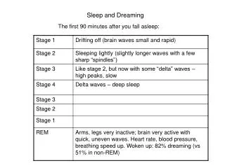
Method Subjects: • 30 right-handed male subjects • Mean age: 22.5 years old (range 20-25 years old) • Subjects were monitored polygraphically over 3 nights spent on a scanner couch, at one-week intervals • Subjects were selected based on those who could maintain two periods of slow wave sleep & 20 minutes of REM sleep during the first two test nights
Experimental Task On the third night, regional cerebral blood flow (rCBF) distribution was recorded during sleep Scans were done when polysomnography showed characteristic sleep patterns (slow-wave & REM sleep) After each sleep scan, the subjects were woken up & asked to describe what was in their minds
Results Positive correlations: Pontine tegmentum Left thalamus Left & right amygdaloid complexes Anterior cingulate cortex Right parietal operculum
Results Negative Correlations: Dorsofrontal prefrontal cortex Parietal cortex Posterior cingulate cortex Precuneus
Discussion • Amygdaloid complexes are involved in the formation & consolidation of memories paired with emotional stimuli • Previous neuroimaging studies suggest that dorsolateral prefrontal areas & precuneus involved in the encoding & retrieval of episodic memory - this study showed that these memory processes are not prominent during REM sleep
Discussion cont’d • Coactivation of anterior cingulate cortex & amygdaloid complexes could account for the emotional & affective aspects of dreams • Other formal characteristics of dreams (temporal distortions, weakening of self-reflective control, amnesia or awakening) might be related to the relative prefrontal deactivation
STRENGTHS Results from the rCBF distribution agree with previous research that suggests there is a functional link between the amygdala, the hippocampal formations & cortical areas during REM sleep These results shed light on the idea that amygdalocortical activation could account for the perceptual components of dreaming This study looked at both positive and negative correlations of PET data
NEXT STEP … Test subjects that vary in age group, gender, handedness - will the same brain regions become activated? Conduct the experiment during the morning or afternoon – will we obtain the same results? Perform the same procedure except this time do not ask participants to stay awake during the preceding second and third night test nights since that obviously produced some fatigue effects in this current study
LIMITATIONS • Only used male subjects – could there be a genetic bias? • Subjects were all right-handed – why not test left-handed subjects? • Subjects were all roughly part of the same age group – could there be an effect of age? • Previous neuroimaging studies have suggested occiptal & temporal areas are activated during REM sleep but that was not shown in this study – WHY? Because group analysis averaged out cortical activations specific to each individual subject or even to each scanned REM sleep episode
THANK YOU FOR LISTENING! Questions…?