Download
1 / 60
620 likes | 992 Vues
Electrical Signals in Animals. Chapter 45. Nerve net. (a) Hydra (cnidarian). Nervous Systems. Consist of circuits of neurons and supporting cells All animals except sponges have some type of nervous system Differ in the way that the neurons are organized into circuits

E N D
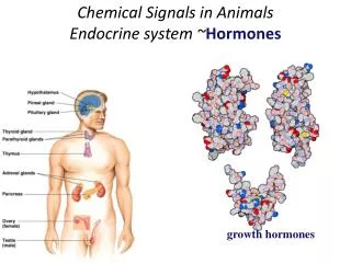
Electrical Signals in Animals Chapter 45
Nerve net (a) Hydra (cnidarian) Nervous Systems • Consist of circuits of neurons and supporting cells • All animals except sponges have some type of nervous system • Differ in the way that the neurons are organized into circuits • The simplest animals with nervous systems, the cnidarians • Have neurons arranged in nerve nets
Radialnerve Nervering (b) Sea star (echinoderm) Nervous Systems • More complex animals contain nerve nets as well as nerves • Bundles of fiber-like extensions of neurons • Sea stars have a nerve net in each arm • Connected by radial nerves to a central nerve ring
Eyespot Brain Nerve cord Transversenerve (c) Planarian (flatworm) Nervous Systems • Cephalization evolved with greater complexity in nervous systems • Clustering of neurons in a brain near the anterior end in worms • Small brain, longitudinal nerve chords constitute the simplest central nervous system
Brain Brain Ventralnerve cord Ventral nervecord Segmentalganglia Segmentalganglion (d) Leech (annelid) (e) Insect (arthropod) Nervous Systems • Annelids and arthropods have more complicated brains • Have segmentally arranged clusters of neurons called ganglia • These ganglia connect to the CNS • And make up a peripheral nervous system (PNS)
Anteriornerve ring Ganglia Brain Longitudinalnerve cords Ganglia (g) Squid (mollusc) (f) Chiton (mollusc) Nervous Systems • Nervous systems in mollusks • Correlate with the animals’ lifestyles • Sessile mollusks have simple systems • While more complex mollusks have more sophisticated systems • Can complete complicated tasks
Brain Sensoryganglion Spinalcord (dorsalnerve cord) (h) Salamander (chordate) Nervous Systems • In vertebrates • The central nervous system consists of a brain and dorsal spinal cord • The PNS connects to the CNS
Neuron Structure • The ability of the neuron to receive and transmit information depends on their structure. Consists of: • Cell body- where most of the organelles are housed • Numerous dendrites- highly branched extensions that receive signals from other neurons • Axon- larger extension that transmits signals, may be covered with a myelin sheath
Dendrite • Receives electrical signals from the axons of adjacent cells. • Axon then sends the signal to the dendrites of other neurons • Cell body, or soma, which includes the nucleus, integrates the incoming signals and generates an outgoing signal in the axon
Dendrites Axon Cell body (c) Motor neuron (b) Interneurons (a) Sensory neuron Neurons • Neurons have a wide variety of shapes • That reflect their input and output interactions • Reflects the number of synapses it has with other neurons
Glia • Glia are supporting cells • That are essential for the structural integrity of the nervous system and for the normal functioning of neurons • In the CNS astrocytes regulate extracellular ion concentrations
Node of Ranvier Layers of myelin Axon Schwann cell Schwann cell Nodes of Ranvier Nucleus of Schwann cell Axon Myelin sheath 0.1 µm • Oligodendrocytes (in the CNS) and Schwann cells (in the PNS) • Are glia that form the myelin sheaths around the axons of many vertebrate neurons • Act as insulators • Multiple Sclerosis is deterioration of the myelin sheath
Resting Potential of a Cell Ion pumps and ion channels maintain the resting potential of a neuron • The resting potential • Is the membrane potential of a neuron that is not transmitting signals • Across its plasma membrane, every cell has a voltage • Called a membrane potential • The inside of a cell is negative • Relative to the outside
Resting Potential of a Cell • The concentration of Na+ is higher in the extracellular fluid than in the cytosol • While the opposite is true for K+ • A neuron that is not transmitting signals • Contains many open K+ channels and fewer open Na+ channels in its plasma membrane • The diffusion of K+ and Na+ through these channels • Leads to a separation of charges across the membrane, producing the resting potential
Gated Ion Channels • Ungated ion channels are always open, results in the resting potential of the cell • Gated ion channels open or close • In response to membrane stretch or the binding of a specific ligand • In response to a change in the membrane potential • Neurons have gated ion channels • Responsible for generating the signals of the nervous system
Starting An Action Potential? • An action potential is a rapid, temporary change in a membrane potential • Has three phases: depolarization, repolarization, and the undershoot • The initial event is a rapid depolarization of the membrane • Membrane potential must shift from its resting potential of –70 mV to about –55 mV
If the threshold potential is reached, channels in the axon membrane open and ions rush into the axon, following their electrochemical gradients • The current flow causes further depolarization • When the membrane potential reaches about +40 mV the membrane experiences a rapid repolarization as ions flow out of the axon
The repolarization event results in the membrane becoming more negative than the resting potential • Called the undershoot • All phases occur in about a millisecond
Regeneration of the Action Potential • An action potential can travel long distances • By regenerating itself along the axon • At the site where the action potential is generated • An electrical current depolarizes the neighboring region of the axon membrane
– – + + + + + + – – + + + + + + Axon Actionpotential An action potential is generated as Na+ flows inward across the membrane at one location. 1 + + – – – – – – – – + + + + + + Na+ – – – – – – + + Actionpotential The depolarization of the action potential spreads to the neighboring region of the membrane, re-initiating the action potential there. To the left of this region, the membrane is repolarizing as K+ flows outward. 2 K+ – – + – – – + – Na+ – – – + + – – – – – + + + + + + K+ Actionpotential The depolarization-repolarization process isrepeated in the next region of the membrane. In this way, local currents of ions across the plasma membrane cause the action potential to be propagated along the length of the axon. 3 K+ – – – – + + + + – + + + + – – – Na+ – – – + + – + + – + + – – – + + K+ Regeneration of the Action Potential
Action Potentials • The speed of an action potential • Increases with the diameter of an axon • In vertebrates, axons are myelinated • Also causing the speed of an action potential to increase • Causes the membranes to have a simulated wider width • Action potentials in myelinated axons • Jump between the nodes of Ranvier in a process called saltatory conduction
Synapses • Neurons communicate with other cells at synapses • In an electrical synapse • Electrical current flows directly from one cell to another via a gap junction • Synchronize the activity of neurons responsible for rapid responses (flight response) • The vast majority of synapses • Are chemical synapses • Not as fast as electrical synapsis
Postsynapticneuron Synapticterminalof presynapticneurons 5 µm Chemical Synapses • In a chemical synapse, a presynaptic neuron • Releases chemical neurotransmitters, which are stored in the synaptic terminal
Synapses • The interface between two neurons is called a synapse. • Just inside the synapse, the axon contains synaptic vesicles that serve as storage sites for neurotransmitters • The sending cell is called the presynaptic neuron and the receiving cell is called the postsynaptic neuron
What Do Neurotransmitters Do? • There are several categories of neurotransmitters • Many neurotransmitters function as ligands that bind to ligand-gated ion channels • Cause the ion channels to open, generating a postsynaptic potential • Causes the start of a new action potential
Central nervous system (CNS) Peripheral nervous system (PNS) Brain Cranial nerves Spinal cord Ganglia outside CNS Spinal nerves The Vertebrate Nervous System • The vertebrate nervous system is regionally specialized • In all vertebrates, the nervous system • Shows a high degree of cephalization and distinct CNS and PNS components
The Central Nervous System • The brain provides the integrative power • That underlies the complex behavior of vertebrates • The spinal cord integrates simple responses to certain kinds of stimuli • And conveys information to and from the brain • The central canal of the spinal cord and the four ventricles of the brain • Are hollow, since they are derived from the dorsal embryonic nerve cord
CNS consists of spinal chord and four ventricles in the brain Contain cerebrospinal fluid Assists in supply of nutrients and hormones to parts of the brain and in removal of wastes Grey matter contains mostly dendrites White matter contains long axons with great bundles of myelin sheaths Gray matter White matter Ventricles The Central Nervous System
The Peripheral Nervous System • The PNS transmits information to and from the CNS and plays a large role in regulating a vertebrate’s movement and internal environment • The cranial nerves originate in the brain • And terminate mostly in organs of the head and upper body • The spinal nerves originate in the spinal cord • And extend to parts of the body below the head
Peripheral nervous system Somatic nervous system Autonomic nervous system Sympathetic division Parasympathetic division Enteric division The Peripheral Nervous System • The PNS can be divided into two functional components • The somatic nervous system and the autonomic nervous system
The Peripheral Nervous System • The somatic nervous system • Carries signals to skeletal muscles • Often considered voluntary • The autonomic nervous system • Regulates the internal environment, in an involuntary manner • Is divided into the sympathetic, parasympathetic, and enteric divisions
Brain structures present in adult Cerebrum (cerebral hemispheres; includes cerebral cortex, white matter, basal nuclei) Diencephalon (thalamus, hypothalamus, epithalamus) Midbrain (part of brainstem) Pons (part of brainstem), cerebellum Medulla oblongata (part of brainstem) Diencephalon: Cerebral hemisphere Hypothalamus Thalamus Pineal gland (part of epithalamus) Brainstem: Midbrain Pons Pituitary gland Medulla oblongata Cerebellum Spinal cord Central canal (c) Adult Brain Structures in an Adult
The brainstem consists of three parts The medulla oblongata, the pons, and the midbrain Parts of The Brain
Brainstem • The medulla oblongata • Contains centers that control several visceral functions • The pons • Also participates in visceral functions • The midbrain • Contains centers for the receipt and integration of several types of sensory information
The Cerebellum • The cerebellum • Is important for coordination and error checking during motor, perceptual, and cognitive functions • The cerebellum • Is also involved in learning and remembering motor skills
Diancephalon • The embryonic diencephalon develops into three adult brain regions • The epithalamus, thalamus, and hypothalamus • The epithalamus • Includes the pineal gland and the choroid plexus • Produce cerebro- spinal fluid
Diancephalon • The thalamus • Is the main input center for sensory information going to the cerebrum and the main output center for motor information leaving the cerebrum • The hypothalamus regulates • Homeostasis • Basic survival behaviors such as feeding, fighting, fleeing, and reproducing