Download
1 / 37
370 likes | 521 Vues
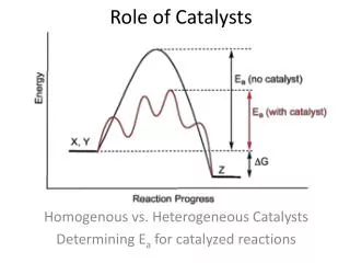
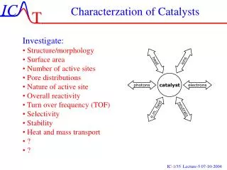
Characterzation of Catalysts. Investigate: Structure/morphology Surface area Number of active sites Pore distributions Nature of active site Overall reactivity Turn over frequency (TOF) Selectivity Stability Heat and mass transport ? ?. The Surface Science Approach.

E N D
Characterzation of Catalysts • Investigate: • Structure/morphology • Surface area • Number of active sites • Pore distributions • Nature of active site • Overall reactivity • Turn over frequency (TOF) • Selectivity • Stability • Heat and mass transport • ? • ?
The Surface Science Approach • Simpler system - Detailed studies • Fundamental insight • Input to catalyst design • The structure gap • The pressure gap • The materials gap Single crystal surfaces as model catalysts.
X-Ray Diffraction (XRD) Bragg ´s Law X-rays in X-rays out catalyst Can be used in situ Gives information on phases and sizes of particles if big enough.
X-Ray Diffraction (XRD) Position reveals crystalline structure Width of peaks reveals particle size 4.5 Å 2.5 Å
X-ray Photoelectron Spectroscopy (XPS) X-rays Photo-electrons catalyst Can be not used in situ as it require vacuum Ekin=hn-EB-F Gives information on elementary coposition and chemical state
Decays following creation of core hole X-ray emission (Electron dispersive x-ray emission - EDX): (hn’)=Eb(K)-Eb(L) L K Auger electron emission: Ekin=Eb(K)-Eb(L1)-Eb(L2) After ~10-14 s L K Photoelectron: Ekin=hn – Eb - F
Surface Sensitive Electrons traveling though a solid has a short mean free path I(z)=I0exp(-z/l)
X-ray Photoelectron Spectroscopy (XPS) Notice how the EB is influenced by chemical environnement
X-ray Photoelectron Spectroscopy (XPS) On combustion catalyst the loading of Pt may be so small that it cannot be detected by XPS (0.1% and 2-5 nm particles).
Extended X-Ray Absorption Fine Structure (EXAFS) catalyst Measure adsorbtion An in situ method Gives information on local atomic distances and coordination number
Electron Microscopy Does not work in situ Gives information on partiles size, shape, composition.
Environnemental EM from Haldor Topsøe A/S Graphite Ni ~1000 Å
Mössbauer spectroscopy hn=14.4 keV DE=10-9 eV An in situ method Rather specialized method since it works only on Fe, Sm,..
Mössbauer spectroscopy example Reveals oxidation state and chemical sourroundings of particular Fe and Mo catalysts
Ion spectroscopy: SIMS Ions out Ions in catalyst SIMS is not an in situ method Is very sensitive for some elements on the ppm level
Ion spectroscopy: LEIS Ions out Ions in catalyst Why a shoulder? Very surface sensitive
Temperature programmed reduction, oxidation and sulfidation Sulfidation in a H2S/H2 mixture
Infrared spectroscopy IR out IR in catalyst IR through An in situ method
Infrared spectroscopy IR is used for identifying intermediates Active sites by adsorption of probe molecules like NO, CO,.. Line position may reveal bonding geometry and bonding strength Surface Science metod is HREELS High Reflection Electron Energy Loss Spectroscopy - photons replaced by electrons
A Surface Science Approach toHeterogeneous Catalysis Natural Gas Ni CH4+H2O CO + 3H2+(CO2) Cu N2+Fe, Ru CH3OH H2 NH3
Low Electron Energy Diffraction (LEED) Requires well-defined surfaces under UHV
LEED picture Reconstruction of a Fe(111) surface into a 5x5 N-Fe(111) surface
LEED Crystal Screen Diffracted beams (-1,1) (0,1) (1,1) (-1,0) (0,0) (1,0) a2* a2 a1* a1 Primary beam (-1,-1) (0,-1) (1,-1)
The Scanning Probe Methods surface z x y current Piezo electric elements tip Requires well-defined surfaces. For STM they must also be conducting
Scanning tunneling microscopy (STM) L. P. Nielsen University of Århus (80Åx80Å) Length scales ”Seeing” atoms Electron tunneling Hardware realisation Applications Basic research
SEM image Crystallites and STM-tip A sharp tip 35 micrometer A. Emundts und H.P. Bonzel
STM Au/Ni(111) Let you sometime, but not always, see the atoms 0.02ML Au 0.8 Ml Au
STM Model Catalyst MoS2 on Au(111) Sulfur removed by atomic hydrogen
Fe/Cu(111) Kanji ”atom” Manipulating atoms CO/Pt(111) ”CO-man” 5 nm Scanned at very low temperatures http://www.almaden.ibm.com/vis/stm/gallery.html
Fe/Cu(111) Quantum Corrals http://www.almaden.ibm.com/vis/stm/gallery.html