Download
1 / 10
100 likes | 104 Vues
Medical problems in dermamatology Inflammatory bowel disease. Cutaneous manifestastions

E N D
Medical problems in dermamatologyInflammatory bowel disease • Cutaneous manifestastions • @ Erythema nodosum, erythema multiformis @ Pyoderma gangrenosa, sweet’s syndrome, other neutrophilic dermatosis @ Small vessel vasculitis, cutaneous polyarthritis nodosa @ Fistula & abscesses in perianal region @ Pruritis ani, vulvi &scrota @ acquired acrodermatitis enteropathica, • Oral associations: • @ Apthous ulcers @ granulomatous chelitis, angular chelitis @ gingival / mucosal swelling @ Cobblestoning of buccal mucosas. • Secondary systemic amyloid may be associated with longstanding inflammatory bowel disease & presents with petichiae & ecchymosis. However, primary macular amyloidosis has nothing to do with inflammatory bowel disease but only present with macular hyperpigmwentation.
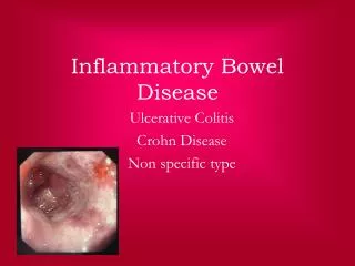
Inflammatory bowel disease (continue) • The main forms of inflammatory bowel disease are Crohn’s disease & ulcerative colitis, The main difference between them is the location& nature of inflammatory changes. Crohn’s disease can affect any part of G.I.T. from mouth to anus, although majority of cases start in the terminal ileum. Ulcerative colitis , in contrast, is restricted to the colon & the rectum.. • Microscopically, ulcerative colitis is restricted to the mucosa ( epithelial lining of the gut), while Crohn’s disease affects the whole bowel wall. • Both diseases may present with vomiting, diarrhea, rectal bleeding, weight loss, severe internal cramps, muscle spasm in region of pelvis & various complaints & diseases like arthritis, pyoderma gangrenosa, cutaneous manifestations, primary sclerosing cholangitis.
Pancreatitis • Cutaneous features: • @ Grey –Turner’s sign: This is a cutaeous sign of acute hemorrhagic pancreatitis & carries grave prognosis, Other causes of Grey- Turner ‘s sign include blunt abdominal trrauma, ruptured aortic aneurysm & ruptured ectopic pregnancy.It is bluish dicolaration OF THE FLANKs
Pancreatitis (Continue) • @Cullen’s sign: Black blue bruising around the umbilicus. • @Pancreatic panniculitis: • Tender fluctant nodules on • the lower legs • @ Jaundice, @ livedo reticularis @ urticaria @ Throbophlebitis migrans: pancreatic malignancy associated
Rheumatoid arthritis • Cutaneous manifestations • @Neutrophilic dermatosis: Pyoderma gangrenosa, rheumatoid neutrophilic dermatoses, sweet’s syndrome, neutrophilic panniculitis; • @ Palisading granuloma: rheumatoid nodules & papules, palisaded neutrophilic & granulomatous dermatitis, interstitial granulomatous dermatitis • @ Vascular: small, medium & large vessel vasculitis, capilaritis, bywater’lesions: • It is a cutaneous manifestation of rheumatoid arthritis. They are cutaneous infarcts related to small vessel rheumatoid vasculitis,. In addition ,they also may present under the nails as painless red brown lesions mimking the splinter hemorrhages of subacute bacterial endocarditis • @ Purpura. • @palmer erythema • @ Thin skin @ Drug reaction to therapy
Palmer erythema • Causes: • @ Familial variant @ Chronic liver disease, alcohol abuse @ pregnancy @ connective tissue disorders e,g. lupus, rheumatoid arthritis, sarcoid @Thyrotoxicosis @ Polycythemia @ Leukemia @ Inflammatory dermatosis e,g. eczema, psoriasis, erythroderma @ Chemotherapy, anti-epileptic merdications @ HTLVI infection @ Paraneoplastic: especially brain malignancies. • Chronic liver disease • Cutaneous feature: • @ Palmer erythema @ Clubbing of the nails, leukoplakia, spider angiomas, @ Diffuse pigmentation @ Loss of axillary & pubic hair @Gynecomastia @ Xanthelasma , if obstructed elements.
Facial flushing • Causes: • @ Physiological: emotional or sexual arousal. @ Menopausal @ Rosacea. @ Carcinoid syndrome @ Hyperthyroidism @ Pheochromocytoma @ Autonomic dysfunction, migraine, neurological lesions @ Systemic mastocytosis @ neoplasm. • Drugs related flushing: • @ ACE inhibitors @ Calcium channel blockers @ Alcohol @ nitrates @ Opiods @ Calcitronin • Carcinod syndrome: • Carcinoid syndrome occurs when a neuroendocrine carcinoid tumor metastasize to the liver & release serotenin & other mediators into the blood streem. Patients may develop a pellagra like photodistributed skin rash & the flushed areas may have violaceous hue. Bronchospasm, diarrhea & cardiac murmur can be seen. The disese is investigated by testing urine for metabolities such as 5-HIAA
Generalized pruritus • Causes: • @ Scabies @ Liver disease @ Malignancy @ Hematological: leukemia, lymphoma, myeloma @ Drug reactions @ Renal failure (late sign) @ Iron, B12 or floater deficiency @ Hyperthyroidism, hypothyroidism @ Psychogenic neuropathy • Lignocaine toxiciity • The maximum safe dose of plain lignocaine in an adult is 3 mg/ Kg, with adrenaline up to 7 mg/kg may be used , In children and the elderly these doses should be halved, !% lignocaine has 1O mg/ml • If a 7O Kg adult may be given up to 7 mg/kg of lignocaine with adrenaline. This equates to 49O n.g. ,or 49 mls of 1% solution
Macular petechial rash • It consists of flattened purple macules less than 3 mm in diameter • Causes: • @ Thrombocytopenia @ Abnormal platelet function disorders @ Scurvy @ venous eczema @ Pigmented purpuric eruption e.g. Schamberg’s • Macular purpura: Larger flat red purple lesions with a diameterr of 5-9 mm. Vasculitis tends to present with palpable purpura.. Vitamin K defeciency results in ecchymoses
Palpable purpuric rash • Causes: • @ Primary vasculitis • @ Immune related vasculitis: idiopathic, sepsis related, drug associated, secondary to connective tissue disorders. • @ Non-vasculitis: Erthema multiformis, PLEVA, pigmented purpuric eruption • Ecchymoses are flat round-oval purpuric lesion often over 1 cm in size, they normally result from minor traumas on a background of dermal fragility, platlet dysfunction or an anticoagulated state.