Download
1 / 1
10 likes | 134 Vues
D9 0µM SG. D9 50µM SG . GAPDH. 38kDa. SEDI. 17kDa. D9 0µM SG. D9 50µM SG. GAPDH. 38kDa. SOD. CD-1 kidney. G37R con L.Sc . [H 2 O 2 ]. 0µM susp . 500µM susp . 1000µM susp . 500µM . 1000µM . 0µM . 17kDa. 38kDa.

E N D
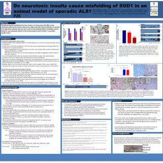
D9 0µM SG D9 50µM SG GAPDH 38kDa SEDI 17kDa D9 0µM SG D9 50µM SG GAPDH 38kDa SOD CD-1 kidney G37R con L.Sc. [H2O2] 0µM susp. 500µM susp. 1000µM susp. 500µM 1000µM 0µM 17kDa 38kDa 17kDa 38kDa 17kDa B) G37R SG G37R Con WT Con WT SG Do neurotoxic insults cause misfolding of SOD1 in an animal model of sporadic ALS? SEDI 17kDa B) A) A) Pierre Zwiegers1, Grace Lee1, Hseuh-Ning. Liu2, Janice Robertson2, & Christopher A. Shaw1 1Neural Dynamics Research Group, Univ. of British Columbia, Vancouver, BC, CAN 2Centr. For Res. In Neurodegenerative Dis., Univ. of Toronto, Toronto, ON, CAN SOD1 ICC P26 B) • Figure 2. Hydrogen peroxide treatment of IMR-32 cells. (A) MTT cell viability shows decrease in cell number with peroxide treatment. (B) Western blot analysis for the SEDI-specific epitope did not show any evidence of SOD1 conformational changes under these conditions. Lanes 1-3: IMR-32 whole cell lysate (15ug/lane; note: significant cell loss evident in lane 3); Lanes 4-6 whole cell lysate (cells in media suspension; 15ug/lane) Lane 7: CD-1 Kidney homogenate (15ug/lane); Lane 8: G37R control lumbar spinal cord homogenate (15ug/lane).Mean +/- SEM shown, One-Way ANOVA; **P<0.001 SEDI ICC [SG] 50µM day3 0µM day6 50µM day6 0µM day9 0µM day3 50µM day9 CD-1 Kidney G37R L.Sc WT Control WT SG G37R SG Having previously established that key features of Guamanian ALS-PDC can be replicated with murine dietary exposure to stigmasterol β-D-glucoside (SG), we wanted to I) determine whether this was due in part to SG-mediated SOD1 misfolding, and II), probe for any synergistic effect on the conformational state of SOD1 in a genetic model of ALS. Objective CD-1 Control G37R Control CD-1 SG Introduction • ALS and the confomational state of SOD1 • Familial ALS (fALS) with mutations linked to the SOD1 genetic locus accounts for approximately 1-2% of • all ALS cases. • Familial forms of ALS are considered to to be clinically and neuropathologically indistinguishable from • sporadic forms of the disorder (sALS). • - SOD1-mediated fALS involves a toxic-gain-of-function with resultant SOD1-protein misfoldingand • aggregation (Rakhit et al, 2007). • - Wild-type and mSOD1 show an increased propensity to misfold and aggregate under conditions • of in vitrooxidation. • A current hypothesis posits that aberrant SOD1 conformational change is the pathobiological • mechanism common to both sporadic and familial forms of ALS (see below). • Recent work conducted challenge this interpretation as development of an antibody selective for the • SOD1Exposed Dimer-Interface (SEDI) is strictly selective for familial forms of the disease and does not • label sALS(Kerman et al, 2010; Liu et al, 2009). • However, Bosco et al, have recently shown that there may be a subset of sALS cases in which the • wild-type SOD1 protein can adopt structural conformations similar to those observed in fALS (2010). • Cycads, sterylglucosides and the etiology of ALS-PDC • Dietary consumption of Cycasmicronesica seeds have been linked to ALS-parkinsonism dementia • complex (ALS-PDC) indigenous to Guam (Whiting, 1963; Borenstein et al, 2007). • -In vivo murine studies using washed cycad flour have shown behavioural and neuropathological • changes similar to ALS-PDC (Wilson et al, 2002). • We have synthesized several water-insoluble sterylglucosides identified in C. micronesicaseeds. • - In vivo and in vitro treatment with sterylglucosides show neural degeneration (Khabazian • et al, 2002 & Tabataet al, 2008). • We have also shown that dietary sterylglucoside exposure in the mSODG37R model results in a more • severe phenotype than observed solely with the underlying genetic defect (Lee et al, 2009 & 2010). A) B) D) C) A) C) Figure 1. SG treatment of IMR-32 cells over a period of 3-9 days. (A) Cell viability as measured by way of MTT assay does indicate an effect of decreased cell viability with SG treatment. However, the mechanism of cell death is unrelated to SOD1 conformational changes as evidenced by (B) Immunocytochemistry (100x; scale bar = 10µm; 1:100 Rb α-SOD1, 1:1000 Rb α-SEDI), and (C) Western blot for the SEDI-specific epitope (1:500 Rb α-SEDI).Lanes 1-6: IMR-32 whole cell lysate (15ug/lane); Lane 7: MW marker; Lane 8: CD-1 Kidney homogenate (15ug/lane); Lane 8: G37R control lumbar spinal cord homogenate (15ug/lane).Mean +/- SEM shown, Mann-Whitney U-test (non-parametric, one-tailed t-test); ***p<0.0001 E) References Acknowledgements Discussion Results Summary Experimental Results Experimental Design • In vivo SG exposure • Both transgenic C57Bl/6 mSODG37R (G37R) and wild-type (WT) littermate animals were • housed in a room maintained at 22°C on a 12/12hr light/dark cycle. • Stigmasterol β-D-glucoside (SG) feeding commenced at 10 weeks of age at 42mg/kilogram of • body weightuntil signs of hind-limb paralysis were evident; control animals were fed control • pellets of equal weight for the duration of the experiment. • - at the endpoint, animals were sacrificed and tissues processed for western blot (WB) or • immunohistochemical (IHC) analysis with an antibody specific for the SEDI epitope (Liu • et al, 2009). • Tissue from CD-1 mice fed SG at 1000 µg/day for 15 weeks were also processed for IHC to • asses the effect(s) on the conformational state of SOD1 in another strain. • In vitro SG exposure • The human-derived IMR-32 neuroblastoma cell line was cultured at 37°C in Eagle’s Essential • Minimum Media, supplemented with 10% (v/v) fetal bovine serum complemented with 1% • (v/v) penicillin/streptomycin. • Stigmasterol β-D-glucoside was solubilized in dimethyl sulfoxide. IMR-32 cells were • exposed to SG diluted in media at a concentration of 50µM according to a treatment • paradigm of 3-9 days. • At the predetermined time points, cells were prepared for the MTT cell viability assay, • Immunocytochemistry (ICC), and Western blot (WB) analysis. • In vitro H2O2exposure • IMR-32 cells were treated with H2O2 diluted in media at concentrations of 500 and 1000µM • according to the following paradigm which was empirically determined to optimally decrease • cell viability: • - Day 1: Cells were treated with H2O2 for 30 min at 37°C, at which point the media was • changed, and cells were allowed to respond to the initial insult overnight in a • humidified incubator. • - Day 2: Cells were re-exposed to H2O2 for 30 min at 37°C. Following the media change, • the cell line was maintained for an additional 6 hours, and the cells were then • prepared for conventional WB and cell viability assays. • Western Blot and immunochemistry • For WB assay : 1° Rbα-SEDI @ 1:500 , Rb α-SOD1 @ 1:2000, Rbα-GAPDH @ 1:5000 • 2° Gt α-Rb HRP @ 1:2000 • For ICC/IHC: 1° Rbα-SEDI @ 1:100 /1:100, Rb α-SOD1 @ 1:100, • 2° Gt α-Rbbiotinylated @ 1:200 Figure 3. (A) Stigmasterolβ-D-glucioside ingestion resulted in motor neuron death as measured by cells labelled for anti-choline acetyltransferase activity (Mean +/- SEM shown; Two-way ANOVA: Genotype: p < 0.0001, Diet: p < 0.05;). Misfolded SOD1 as labeled by the SOD1 Exposed Dimer Interface (SEDI) antibody is evident only in transgenic animals engineered to express mutated human SOD. SG feeding did not induce or exacerbate the SOD1 conformational changes as assayed via (B+C) Western blot (Mean +/- SEM shown, One- way ANOVA ***p<0.0001) or E) immunohistochemistry.Similarly (D), SG toxicity was not found to involve a pathway indicative of SOD1 misfolding in CD-1 animals exposed to the toxin. (scale bar = 10µm; Top panel: Rbα-SEDI @ 1:1000 in the spinal cord of CD-1 mice, Lower panel: Rbα-SEDI @ 1:100; except G37R Con @ 1:1000 in the spinal cord of C5Bl/6 mSODG37R or wild-type animals). WT SG G37R SG WT CON G37R Con • Stigmasterol β-D-glucoside neurotoxicity under these specific experimental • conditions does not induce or exacerbate SOD1 conformational changes as • identified by an antibody specific for the SOD1 Exposed Dimer-Interface • (SEDI) epitope. • - These results are indicative that SG-induced neurotoxicity is similar to • some forms of sALS in that misfolded SOD1 (as identified through • anti-SEDI labelling) is not detected (Liu et al, 2009). • The in vivo and in vitro models outlined here may be used to further • understand the mechanism(s) of disease progression in neurodegenerative • conditions exacerbated by environmental agents with or without an • inherent genetic predisposition. • Future studies would benefit by exploring the conformational state of • SOD1 with the use of antibodies specific for a myriad of epitopes • indicative of the misfoldedand/or oxidized protein. • Prolonged SG treatment of IMR-32 cells expressing endogenous wild-type human SOD exhibited cell loss but failed to induce SOD1 misfolding as labelled with an antibody specific for the SEDI epitope. • Hydrogen peroxide-induced oxidation failed to show evidence of the exposed SEDI epitope even with a demonstrated decrease in cell viability. • Chronic dietary SG exposure in mice did not induce SOD1 misfolding and failed to demonstrate a synergistic interaction with the G37R mutation on the conformational state of SOD1 . Borenstein, A.R., Mortimer, J.A., Schofield, E., Wu, Y., Salmon, D.P., Gamst, A., Olichey,J., Thal, L.J., Silbert, L., and Kaye, J. (2007). Cycad exposure and risk of dementia, MCI, and PDC in the Chamorro population of Guam. Neurology 68: 1764-71. Bosco, D.A., Morfini, G., Karabacak, N.M., Song, Y., Gros-Louis, F., Pasinelli, P., Goolsby, H., Fontaine, B.A., Lemay, N., Mckenna-Yasek, D., Frosch, M.P., Agar, J.N., Julien, JP., Brady, S.T., & Brown, R.H. Wild-type and mutant SOD1 share an aberrant conformation and a common pathogenic pathway in ALS. (2010). Nature Neuroscience, 13(11): 1396-1403. Kerman, A., Liu, HN., Croul, S., Bilbao, J., Rogaeva, E., Zinman, L., Robertson, J. & Chakrabartty, A. (2010). Amyotrophic lateral sclerosis is a non-amyloid disease in which extensive misfolding of SOD1 is unique to the familial form. ActaNeuropathologica, 119: 335-344. Khabazian, I., Bains, J. S., Williams, D. E., Cheung, J., Wilson, J. M., Pasqualotto, B. A., Pelech, S. L., Andersen, R. J., Wang, Y. T., Liu, L., Nagai, A., Kim, S. U., Craig, U. K., & Shaw, C. A. (2002). Isolation of various forms of sterol beta-d-glucoside from the seed of Cycascircinalis: Neurotoxicity and implications for ALS-parkinsonism dementia complex. Journal of Neurochemistry, 82, 516–528. Lee, G., Hilton, B.J., & Shaw, C.A. (2009). Exposure to sterylglucosides in vivo contributes to disease progression in the mSODG37R line 29 mouse model of ALS. Amytrophic Lateral Sclerosis, 10 (s1): 84-96. Lee, G., Lam, O.T.H., and Shaw C.A. (2010). Determination of disease progression with early exposure to sterylglucosides in the mSODG93A mouse model of amyotrophic lateral sclerosis. SFN 2010. Liu, HN., Sanelli, T., Horne, P., Pioro, E.P., Strong, M.J., Rogaeva, E., Bilboa, J., Zinman,L., & Robertson, J. (2009). Lack of evidence of monomer/misfolded superoxide dismutase-1 in sporadic amyotrophic lateral sclerosis. Annals of Neurology, 66(1): 75-80. Rakhit, R., Roberston,J., VandeVelde, C., Horne, P., Ruth, D.M., Griffin,J., Cleveland, D.W., Cashman, N.R., & Chakrabartty, A. (2007). An immunological epitope selective for pathological monomer-misfolded SOD1 in ALS. Nature Medicine, 13(6): 754-759. Tabata R.C., Wilson J.M.B., Ly P.,Zwiegers P., Kwok D., Van Kampen J.M., Cashman N., and Shaw C A. (2008). Chronic exposure to dietary sterol glucosides is neurotoxic to motor neurons and induces an ALS-PDC phenotype. Neuromolecular Medicine, 10(1): 24-39. Whiting M.G. Toxicity of Cycads. (1963). Economic Botany 17: 271-302. Whiting, M.G. Food practices in ALS foci in Japan, the Mariana, and New Guinea. (1964). Fed.Proc. 23: 1343-1345 . Wilson, J. M., Khabazian, I., Wong, M. C., Seyedalikhani, A., Bains, J. S., Pasqualotto, B. A., Williams, D. E., Andersen, R. J., Simpson, R. J., Smith, R., Craig, U. K., Kurland, L. T., and Shaw, C. A. (2002). Behavioral and neurological correlates of ALS- parkinsonism dementia complex in adult mice fed washed cycad flour. Neuromolecular Medicine, 1, 207–221. This work has been made possible by grants from NIH (Shaw), ALS Society of Canada (Shaw, Robertson), the American ALS association (Robertson), the US Muscular Dystrophy Association (Robertson), the CIHR Neuromuscular Research Partnership Program (Robertson) and the Scottish Rite Charitable Foundation of Canada (Shaw). Neural Dynamics Research Group