Download
1 / 28
290 likes | 320 Vues
Understand anatomy basics including positions, planes, terms, body regions, movement, and the skeletal system. Learn about bone functions and classifications. Video link included.

E N D
FIRST LECTURE Introduction to Anatomy ANATOMICAL TERMS AND SKELETAL SYSTEM هذا العمل لا يغني عن المصادر المطلوبة
SKELETAL SYSTEM:BONE • Objectives: • Define the word “ANATOMY” • Enumerate the different anatomical fields • Describe the anatomical position • Describe different anatomical terms of position and movements as well different anatomical planes • Classify bones according to shape , structure and development • Enumerate different bones of both axial and appendicular skeleton
What is Anatomy? It’s the Study that deals with the structure and shape of the Body parts & their relationships to one another.
ANATOMICAL SCIENCES • Gross Anatomy : The study of human body with a naked eyes • Microscopic Anatomy ( Histology ) : Study of fine structure (cell and tissue ) of the human body with the help of microscope • Developmental Anatomy ( Embryology ) • Radiological Anatomy : Study of bodily structure using radiograph and other imaging methods • Applied Anatomy: The practical application knowledge to diagnosis and treatment • Surgical Anatomy: Study of anatomical structure in reference to the surgical diagnosis and treatment • Cross sectional Anatomy: Studying the humane body through a transverse cut through a structure or tissue
Skeletal System Includes: • 1.Bones • 2.Joints(Articulations between bones)
Anatomical Terminology : “To prevent misunderstanding a special set of terms are used to describe identification and location of body structures”. Anatomical position : The standard position in which the body assume to describe its parts . • This position has 4 features : 1- Body is erect 2- Feet parallel 3- Palm facing forward 4- Arms hanging by the side
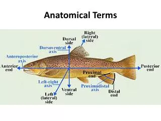
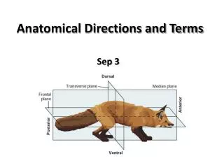
Terms of Position: • Superior (cranial ,rostral ): near to head . Inferior (Caudal): away from head. • Anterior ( ventral ): near to front . Posterior (dorsal) : near to back. • Medial : near to median plane Lateral : away from median plane • Proximal : near to trunk Distal : away from trunk • Superficial : near to skin (surface) Deep : away from skin *Intermediate: the relative location of an anatomical structure lying between two other structures:
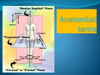
ANATOMICAL PLANES & SECTIONS • Sagittal (median): - A cut made along a longitudinal plane dividing the body into 2 equal halves (right & left). - The plane passing through the midline of the body, cutting the body into the right and left equal halves is called a midsagittalormedian plane. • Parasagittal (paramedian): - divides the body into 2 unequalparts (right & left). • Frontal (coronal): - A cut made along a longitudinalplane. - divides the body into anterior&posteriorparts • Transverse (cross): - A cut made along a horizontal plane. - divides the body into superior & inferiorparts.
Terms of Regions: • Cranial (cephalic) • Cervical • Thoracic • Abdominal • Pelvic • Plantar • Palmar
Body Cavities • There is two sets of internal cavities which areventraland dorsal. • Ventral Body Cavity: divided by the diaphragm into: • 1. Thoracic Cavity: superiorto diaphragm (above the diaphragm),contains heart & lungs. • 2. Abdominal cavity: inferiorto diaphragm (below the diaphragm)contains stomach, intestine, liver, urinary bladder, etc… • Dorsal body cavity: divided into 2 parts continuouswith each other: • 1. Cranial cavity: space inside skull, contains brain. • 2. Spinal cavity: space inside vertebral column, contains spinal cord.
Abdominopelvic Regions • The Abdominopelvic area is divided into 9 regions by 2 vertical & 2 horizontal lines or planes. • Objective: To locate the different organs in each region.
TERMS OF MOVEMENT 1-3 • Flexion: approximation of 2 parts(decreasing the angle between 2 parts) • Extension: straightening (increasing the angle between 2 parts) • Abduction: away from median plane • Adduction:towards median plane • Lateral rotation: rotation away frommedian plane • Medial rotation: rotation toward median plane • Circumduction: combined movementsof flexion, extension, abduction &
TERMS OF MOVEMENT 2-3 -Opposition: bringing tips of fingers and thumb together as in picking something up -Supination: - Lateral rotation of the forearm. - The palm faces Anteriorly. • The radius and ulna are Parallel. • Pronation: - Medial rotation of the forearm. - The palm faces Posteriorly - The radius Crosses the ulna and the two bones form an X.
TERMS OF MOVEMENT 3-3 Planterflexion: - Depressing the foot (down ). • Movement with pointing the toes. Dorsiflexion: - Up movement of the foot • (Standing on the heels) Inversion: The sole faces in a Medial direction. Eversion: The sole faces in a Lateral direction. https://youtu.be/5YcNAPzDxDg
Functions of bone e.g.: Calcium and phosphorus
Classification on bone • Bones are classified based on three things: 1- Shape: Long, Short, Irregular , flat 2- Structure: Compact, Spongy 3- Development: Membrane, Cartilage
The Skeleton The skeleton is formed from 206 Bones
Bones of the axial skeleton The Skull Formed from two sets of bones: • 1. Cranium(bones enclosing and covering the brain): Frontal, Temporal, Occipital, Parietal, Sphenoid • 2. Facial bones (bones that form the skeleton of the face): Maxilla, Nasal, Zygomatic, Mandible
VERTEBRAL COLUMN • It is a flexible curved structure formed of 33 vertebrae (irregular bones) • Running through its cavity is the spinal cord • Formed of : Cervical vertebrae 7 Thoracic vertebrae 12 Lumbar vertebrae 5 Sacral vertebrae fused to form sacrum (triangular bone) 5 coccygeal vertebrae fused to form coccyx (small bone)4
Sternum (Flat bone) : Has 3 parts : Manubrium Body xiphoid process Ribs : • Number: vertebrae 12 pairs • All ribs articulate with vertebrae • Only upper 7 pairs articulate with sternum • The first 7 are called true ribs • The ribs 8,9 and 10 are called False ribs • The ribs 11 and 12 are called Floating ribs
BONES OF APPENDICULAR SKELETON PECTORAL GIRDLE • Connects upper limb with axial skeleton • Formed of: clavicle & scapula(2 bones on each side) PELVIC GIRDLE • Connects lower limb with axial skeleton • Formed of: hip bone (one bone on each side)
BONES OF APPENDICULAR SKELETON UPPER LIMB • Bone of arm: humerus • Bones of forearm: radius(lateral) & ulna (medial) • Bones of hand: • 8 carpal bones • 5 metacarpal bones • 14 phalanges: 2 for the thumb & 3 for each of medial 4 fingers
BONES OF APPENDICULAR SKELETON Lower limp Bones of thigh Femur . Bones of leg Fibula (lateral) & Tibia(medial). Patella. Bones of foot : 7tarsalbones . 5metatarsalbones . 14phalanges : 2for big toe & 3for each four lateral toes .
LONG BONES metaphysis (The region between diaphysis and epiphysis) contains thin plate of cartilage called theepiphyseal plate that is responsible for the lengthwise growth of the long bones. Diaphysis(shaft) -long & cylindrical - Compact bone - Covered on its external surface by a fibrous connective tissue membrane called the periosteum. - Has a cavity called the marrow cavity. In adults, the marrow cavity is a storage area for fatand contains yellow marrow.In infants, it contains red marrow and is the site of blood cells formation
Periosteum • Role of Periosteum: • Protects the bone • Gives attachment to muscles • Carries blood vessels and nerves to bone • Deposits new bone on the surface thus increases the girth of bone
LONG BONES Epiphysis (The two ends) - Spongy Bone - lined by a thin layer of compact bone - Its external surface is covered by a layer of hyaline cartilage called the articular cartilage (provides smooth slippery surface that decreases friction at joint surfaces) *Growth of bone Increase in length: epiphyseal plates Increase in girth: periosteum*
Some links to learn more: • Videos: • https://www.youtube.com/watch?v=rDGqkMHPDqE&feature=youtu.be • https://www.youtube.com/watch?v=uBGl2BujkPQ • https://www.khanacademy.org/science/health-and-medicine/human-anatomy-and-physiology/skeletal-system/v/skeletal-structure-and-function • https://www.youtube.com/watch?v=TnY6l9hMOew&feature=youtu.be • Online quiz: • https://www.onlineexambuilder.com/anatomical-terminology-skeletal-system/exam-35333 • https://www.onlinequizcreator.com/anatomical-terms-and-skeletal-system/quiz-208453
Credits: Team members: Team leaders: Abdullah Jammah Abdulmalik Alhadlaq Majed Alzain Yazeed Alsuhaibani Rakan Bahammam Mosaed Alnowaiser Khalid Aleedan Mohammed Alyousef Mohammed Nasr Mohammed Ghandour Dania Alkelabi Heba Alnasser Deena Alnowiser Jawaher Alkhayyal Rana Barasain Wejdan Alzaid Shouq Albogami Lara Alsaleem Ghadah Almazrou Ameera NIazi Lama Alfozan Nawaf Alkhudairy Jawaher Abanumy